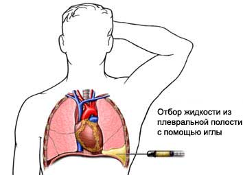
Plevrotsentez – Aspiration plevralynoy zhidkosti
Description thoracentesis
Pleurisy – fluid accumulation in the space between the lungs and the chest wall. This area is called the pleural cavity. Thoracentesis is a procedure to remove fluid from this area.
There are two types of thoracentesis:
- Therapeutic thoracentesis – It serves to reduce the symptoms of fluid accumulation;
- Diagnostic thoracentesis – to determine the cause of fluid accumulation.
Reasons for thoracentesis
The pleural cavity is always a small amount of liquid. When this space too much fluid accumulates, it can obstruct breathing.
The doctor can examine the extracted liquid podadobitsya. Accumulation of fluid can be a symptom of certain diseases or disorders:
- Congestive heart failure (CHF);
- Pulmonary infections;
- Kidney disease;
- Pulmonary embolism (a blood clot in the lungs);
- Cancer;
- Liver disease.
Factors, that may increase the risk of complications:
- Smoking.
Possible complications of thoracentesis
Complications are rare, but the procedure does not guarantee the absence of risk. If you plan to thoracentesis, you need to know about possible complications, which may include:
- Collapsed lung;
- The new fluid accumulation;
- Bleeding;
- Infection;
- Damage to the liver and spleen.
Factors, that may increase the risk of complications:
- Previous lung surgery;
- Long-term, irreversible lung disease (such as emphysema or asthma);
- Factors, affecting the normal blood clotting.
How is thoracentesis?
Prepare for thoracentesis
Your doctor may prescribe:
- A full medical examination;
- Roentgen – test, which uses X-rays to take pictures of organs inside the body;
- CT scan – such as X-rays, which uses computer, to make pictures of structures inside the body;
- Ultrasound examination – uses sound waves, to make pictures of structures inside the body;
- Blood tests.
Anesthesia
It will use local anesthesia, that numb the area of the needle.
Procedure thoracentesis
You, usually, You must sit up straight on the edge of the bed or chair. Hands are on the table. The doctor may use ultrasound, to determine the area of the accumulation of pleural fluid. It will be sterilized by a small patch of skin on the back, breast or under the arm. For anesthesia anesthesia is used. The needle is inserted into the pleural cavity between the ribs. Sometimes, it may additionally introduced a thin catheter. You must avoid coughing, deep breathing or movements during thoracentesis. The syringe will collect some or all of the liquid.

How long will thoracentesis?
About 15 minutes.
Plevrotsentez – Will it hurt?
You may feel some pain or burning while the needle. When the liquid is shown in, you can feel the tension. If there is severe pain, shortness of breath, or fainting, Tell your doctor or nurse.
Care after thoracentesis
Care in a hospital
If thoracentesis is performed to diagnose, fluid will be sent to the laboratory for testing. Often, after the procedure is performed chest X-ray, to ensure that the liquid has been removed and there are no signs of lung collapse.
Home Care
Keep the skin area, where the needle was inserted, clean and dry. To ensure the normal recovery, Be sure to follow your doctor's instructions.
If you perform diagnostic thoracentesis, Ask the doctor, when to expect the results.
Contact your doctor after thoracentesis
Upon returning home after thoracentesis should see a doctor, If the following symptoms:
- Signs of infection, including fever and chills;
- Redness, edema, strong pain, bleeding, or discharge from the needle insertion site;
- Pain, which does not pass after taking pain medication appointed;
- Cough, shortness of breath or chest pain;
- Coughing up blood;
- Pain with a deep breath.
