Ultrasound of the heart – Echocardiogram
Description ultrasound of the heart
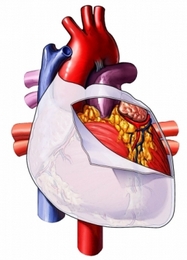
An echocardiogram uses sound waves (ultrasound), to examine the size, shape and motion of the heart.

The test shows:
- Four chambers of the heart;
- Heart valves and the walls hearts;
- Blood vessels entering and leaving the heart;
- Bag, that surrounds the heart.
In addition to the standard test, There are specialized methods of echocardiograms:
- Contrast echocardiogram – injected into a vein in a special solution, helps to see clearly the heart;
- Stress echocardiography – record activity of the heart during the cardiac stress test;
- Echocardiogram with Doppler ultrasound – It helps your doctor to evaluate blood flow;
- Transesophageal echocardiography – to provide a clear view of the heart, ultrasound device is put down your throat. Doctor, perhaps, You need to use this procedure to, to see part of the heart, Poor visibility from other angles;
- Besides, if you have following diseases, you may need this test, rather than the standard echocardiogram:
- Some lung diseases;
- Obesity.
- Besides, if you have following diseases, you may need this test, rather than the standard echocardiogram:
Causes of heart ultrasound
Echocardiography can be carried out as follows:
- Assessment of heart murmurs;
- Diagnosis of diseases of the heart valve;
- Search changes in the structure of the heart;
- Motion estimation of the chamber walls and damage to the heart muscle after a heart attack;
- Evaluation of the various parts of the heart in people with chronic heart disease;
- Determination of fluid accumulation around the heart;
- Determination of proliferation of heart;
- Assessment and monitoring of congenital malformations;
- Checking blood flow through the heart;
- Assessment of damage to the heart and blood vessels from injury;
- Test heart function and diagnose problems of the heart and lungs;
- Evaluation of chest pain;
- Search blood clots in the heart chambers.
How is an echocardiogram?
Preparation for ultrasound of the heart
They can be assigned to the following procedures:
- Medical checkup;
- Electrocardiogram – test, which detects heart activity by measurement of electrical current through the heart muscle.
Description ultrasound of the heart
On special gel is applied to the chest, which helps spread the ultrasonic waves. The specialist applies a small hand-held device (the so-called sensor) to the skin. The transducer sends sound waves toward the heart. The sound waves are then reflected back, captured and converted into electrical impulses. These pulses are displayed as an image on the screen.
The technician can make static pictures or video recording movements of the heart. To get a clear and complete picture, you may need to bring the sensor to different areas of your chest. You may be asked to change position and breathe slowly, breathe out, or hold your breath.
After the ultrasound of the heart
Chest will be cleared from the gel.
How long will the ultrasound of the heart?
30-60 minutes.
Ultrasound of the heart – Will it hurt?
No.
Explanation ultrasound of the heart
The images are analyzed by experts. Based on these data the doctor will recommend treatment and further testing.
Call your doctor
After the ultrasound of the heart, call your doctor, if there was a deterioration of cardiovascular symptoms.
