Myelography – Myelogram
Description myelography
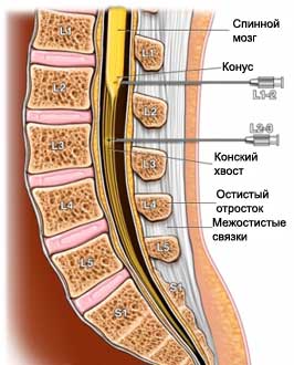
Myelogram – X-ray test, which uses a special radiopaque substance to take pictures of the spinal cord. Together with the X-ray, contrast agent can help the physician to clearly define the space, which contains the spinal cord and nerves. Roentgen – test, which uses X-rays, to take pictures of structures inside the body.
Reasons for myelography
The procedure is used to diagnose problems of the spinal cord, such as:
- Spinal cord growth;
- Herniated disk;
- The narrowing of the spinal canal.
Possible complications of myelography
Complications are rare, but the procedure does not guarantee the absence of risk. If you plan to myelography, you need to know about possible complications, which may include:
- Headache;
- Allergic reaction to the contrast dye;
- Bleeding;
- Inflammation or infection of the spinal cord.
How is myelography?
Preparation for the procedure
Your doctor may prescribe or to hold the next:
- Survey and study of history;
- Ask about the availability of pregnancy – myelography is not typically done for women, who are pregnant;
- Revise list of drugs taken. Maybe, You have to stop taking or change the dosage of some drugs;
- Determine, Do you have any allergies;
- Maybe, prescribe a mild sedative, to help you relax.
In anticipation of the myelography:
- The night before, you can not eat solid food. You can continue to drink fluids;
- If your doctor prescribes a sedative:
- It is necessary to arrange a ride home after surgery. Besides, We need to organize assistance in the home;
- Take the sedative before myelography, in accordance with the instructions of the doctor.
Anesthesia
Myelography, usually, It is carried out without anesthesia. Your doctor may give you a mild sedative. Sometimes a local anesthetic is applied, to reduce the pain of needle insertion.
Procedure myelography
You will lie on your side or face down. You can also sit on the edge of the couch, leaning forward. In the back can be introduced a local anesthetic injection.
A doctor inserts a needle into the space between the vertebrae. From the spinal canal is removed a small amount of liquid. Further, the needle will be inserted through the radiopaque substance. The doctor will use a method of medical imaging called fluoroscopy – a combination of X-ray apparatus with a camera and a screen.
For, to take pictures, you need to lie on your stomach on a special table. You will be secured in a fixed position. The table will be rotated, then the doctor will perform shots back. In some cases, while taking pictures, you have to hold your breath. You may be asked to turn slightly to one, and then in the opposite direction.
Quite often, after the doctor performs myelography computed tomography, that allows you to see the spread of the contrast agent.

Immediately after myelography
You may be asked to wait in the hospital, until the doctor will examine pictures. It may take about an hour.
How long will myelography?
About 30-60 minutes (CT scan will take on 30-60 minutes longer).
Myelography – Will it hurt?
It may feel some pressure or pain when the needle is inserted.
Care after myelography
- If you took a sedative, you can not get behind the wheel, operate machinery, or make important decisions, until the sedative wears off;
- Avoid strenuous exercise (including bends) during 1-2 days.
Contact your doctor after myelography
Returning home, you need to see a doctor, If the following symptoms:
- Signs of infection, including fever and chills;
- Headache, which lasted more than 24 hours;
- Excessive nausea or vomiting;
- Stiff neck;
- Numbness in your legs;
- Problems with urination or bowel;
- Symptoms of an allergic reaction (eg, hives, itch, nausea, swelling and itching of the eye, swelling of the throat, labored breathing);
- The deterioration of the current symptoms.
