Elimination of atrial septal defect in a child – transkateternaya procedure
Description of the operation to eliminate atrial septal defect in a child
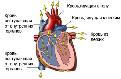
Atrial septal defect – opening in the wall between the two upper chambers (right and left atrium) hearts. Transkateternaя procedure – minimally invasive way to close the hole. During this procedure a special device is implanted, which allows you to close the opening. As the child recovers the device causes heart tissue to grow and more fully close the hole.

Reasons for surgery to remove atrial septal defect in a child
If a child is born with a hole between the upper chambers of the heart, blood can flow back, the right side of the heart and the lungs. It causes the heart to work harder. Over time, this can cause damage to the blood vessels in the lungs and congestive heart failure. The operation is performed, to close the opening.
Most children this opertsii causes a significant improvement in cardiac function.
Possible complications in eliminating atrial septal defect in a child
Complications are rare, but no operation ensures no risk. Before, how to perform the operation, you need to know about possible complications, which may include:
- Bleeding at the site of catheter insertion;
- Damage to the arteries;
- Allergic reaction to x-ray dye;
- The formation of a blood clot (thrombus);
- Infection, including endocarditis (inflammation of the lining of the heart muscle);
- Reaction to anesthesia (eg, dizziness, lowering blood pressure, breathlessness);
- Arrhythmia (abnormal heart rhythm).
Some factors, that may increase the risk of complications:
- Existing diseases (eg, coagulation failure, kidney problems);
- Recent infection.
How is the elimination of atrial septal defect in a child?
Preparing for Surgery
The doctor examines the child and, if necessary, may appoint the following tests:
- Blood and urine tests;
- Echocardiogram – test, which uses sound waves to visualize functioning of the heart;
- Electrocardiogram (ECG ) – test, that records heart activity by measuring electrical current, passing through the heart muscle;
- Chest X-ray – test, which uses X-rays, to take a picture of structures inside the chest;
If a child needs to stop taking certain medications, the doctor will tell about it. It is necessary to ask the doctor, how long before the surgery the child must stop eating and drinking.
Anesthesia
The operation is performed under general anesthesia. The drug is administered through an IV in the arm or hand. During the operation, the patient is asleep.
Description transcatheter surgery to remove atrial septal defect in a child
Through a vein in the arm the child entered the necessary fluids and medications. Catheter (special tube) It is inserted in the arm or groin. After that will be placed on the chest electrodes, that let you capture an electrocardiogram and monitor heart activity.
The catheter is inserted into a blood vessel in the heart and progresses. Radiopaque dye is introduced, so the doctor can see the heart on X-rays. Also for these purposes may be used echocardiogram. Before closing the hole, the doctor must know its size. To do this, a balloon catheter is guided into the upper chambers of the heart. The balloon is inflated and allows to measure the hole.
When the doctor knows the size of the defect, near one directed catheter with a device for closing the opening. There are various types of such devices. Some of them are able to close the hole on both sides. Other devices open like an umbrella. Once the device is mounted in place, the doctor removes the catheter. At the site of insertion of the catheter is placed bandage.
Immediately after surgery
A child is placed for the monitoring in the NICU (OBE) to monitor vital signs. In addition, the child needs some time to lie, and staff should change his bandage in place the catheter insertion.
How long will the surgery?
1-2 o'clock
Will it hurt?
If pain or soreness during recovery will be appointed painkillers.
The average hospital stay
Typically, the duration of stay of 2-4 day. In some cases, a child can be discharged the next day after surgery. If complications arise, the doctor can extend the term of at the hospital.
Postoperative care
In the hospital
The hospital staff does the following:
- Carry out an analysis (blood, Urine, ECG, etc.);
- It gives painkillers;
- Gradually transfer the child to a normal diet.
It is necessary to ask the child to lie still for several hours. This is to prevent bleeding. For the estimation of the radiopaque dye is necessary to drink plenty of fluids.
Nursing homes
When the child returns home, you need to do the following:
- If prescribed by a doctor, the child is given antibiotics. This will help prevent endocarditis;
- As necessary, provided the child painkillers. To alleviate the discomfort, to the place of insertion of the catheter Apply ice;
- It is necessary to drink plenty of fluids, to completely withdraw from the body radiopaque dye;
- The child may be at risk of blood clots in blood vessels. As directed by the physician should be given medicine to prevent blood clots;
- Gradually, the need to transfer the baby to the daily diet;
- A child needs a rest for a few days after the operation. It is not recommended at this time to play energetic games.
It is necessary to go to the hospital in the following cases
- There are signs of infection, including fever and chills;
- Increased perspiration;
- Redness, edema, strong pain, bleeding or discharge from the catheter site;
- Nausea and / or vomiting;
- Increased pain;
- Problems with urination (eg, pain, burning, frequent urination, blood in urine) or inability to urinate;
- Cough, shortness of breath or chest pain;
- Fatigue;
- Rash;
- Reluctance to eat and drink.
It is necessary to call an ambulance in the following cases
- Fast breathing or trouble breathing;
- Blue or gray skin color;
- The child does not wake up or does not respond to treatment;
- Severe chest pain;
- Cardiopalmus;
- Weakness or fainting;
- Signs of stroke (eg, drooping facial muscles, blurred vision or speech, difficulty walking).
