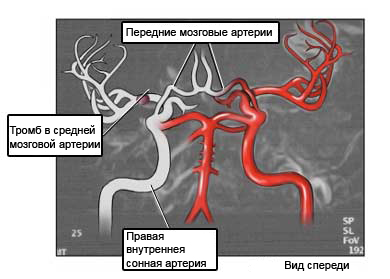
Magnetic resonance angiography – MRA
Description of magnetic resonance angiography
Magnetic resonance angiography is an examination of the blood vessels using magnetic resonance imaging (MRT). Using a large magnet, radiowave transmitter and computer, using MPA can see two-dimensional and three-dimensional images of blood vessels.

Reasons for magnetic resonance angiography
This procedure is carried out to:
- Identify patients, restricted, extended, or blocked blood vessels;
- Find internal bleeding.
How is magnetic resonance angiography?
Preparation for the procedure
If your doctor prescribes a sedative before the procedure:
- You can not eat or drink anything for at least four hours before analysis;
- For 1-2 hours prior to analysis is necessary to take a sedative.
The MRI center:
- The patient will be asked about the following:
- Previous operations;
- Pregnancy;
- Allergies;
- The presence of any metal objects in the body;
- Also ask about the presence of objects in the body, that would interfere with MRI, such as:
- Pacemaker or implantable defibrillator;
- Nerve stimulator;
- Ear implants;
- Metal fragments in eyes or in any other part of the body. You must inform your doctor, if the work involves metal filings or particles;
- Implanted device, eg, insulinovaya pump;
- Metal plate, studs, Screws, or surgical staples;
- Metal clips from aneurysm surgery;
- Bullets;
- Any other large metal objects in the body (dental fillings and braces properly maintained MRI procedure);
- It is necessary to remove all metal objects (eg, jewelry, Hearing Aids, glasses);
- X-rays can be performed, to check for any metal objects in the body.
Before the procedure
- Will be given earplugs or headphones (MRI machine makes a loud noise);
- Maybe an injection of contrast material.
Procedure magnetic resonance angiography
If you are using a contrast agent, hand inserts a thin needle, before being placed in the MRI apparatus. The contrast will be introduced during one of a series of photos. This helps to better see some organs and blood vessels.. Rarely can be an allergic reaction to the contrast dye.
Patients lie on a special table. This table is moved to the MRI machine. MRI analysis will consist of 2-6 implementation procedures photos. The interval between treatments 2-15 minutes. At run time, you must lie still photography. Maybe, You will need to hold your breath. The operator will be in another room. The patient is able to talk to him through the intercom.
After the procedure
- Examinees are asked to wait in the MRI machine, It is considered to be made photos. Technique, perhaps, need to make additional image.
- If you have made a sedative, you can not drive a car or use machines, until after sedation;
- If the examinee – nursing mother, which introduced the contrast dye, you need to find out from the doctor, when you can resume breastfeeding. Studies have found no side effects a child, if a nursing mother has been introduced contrasting paint;
- It is necessary to follow the doctor's instructions.
Duration of treatment
The survey covers 40-90 minutes.
Will it hurt?
Examination is painless. Nonetheless, maybe next:
- Loud noise from the operating unit MRI;
- Burning, when the needle is inserted into a vein (If you are using a contrast agent).
The results of magnetic resonance angiography
The doctor examines the photos and prescribe the necessary treatment.
It is necessary to go to the hospital in the following cases
- Worsening of symptoms;
- Allergic symptoms (It has been used the contrast agent).
