Anemia, due to a deficiency of cyanocobalamin - Vitamin B12
Anemia, caused by deficiency of the enzyme kobamidnogo (B12-cofactors) methylcobalamin characterized by the appearance in the bone marrow megaloblasts, intramedullary destruction erythrokaryocytes, perhromnoy megalocytic anemia, thrombocytopenia, lejko- and neutropenia.
With a deficit of coenzyme second - dezoksiadenozilkobalamina appear atrophic changes in the mucous membrane of the stomach and intestines, and changes in the nervous system as a funicular myelosis.
The etiology and pathogenesis of anemia, due to a deficiency of cyanocobalamin
Cyanocobalamin is involved in humans in two important reactions. First of all, it methylcobalamin coenzyme required for the synthesis of thymidine, which is part of the DNA. This reaction provides the expansion of hematopoietic cells and cells of the mucous membrane of the stomach and intestines.
The second reaction, which involved coenzyme cyanocobalamin dezoksiadenozilkobalamin - is decay and synthesis of certain fatty acids. Violation of the synthesis of fatty acids reduces the synthesis of myelin, and violation of their decay - to the accumulation in the body toxic to nerve cells methyl malonate. The first reaction is carried out through folic acid: methylcobalamin is formed with the participation of the active form of folic acid (5,10-methylene-tetragidrofolievaya Chisloth), necessary for the synthesis of thymidine. For the second reaction, folic acid is required. If patients with impaired cobalamin doctor prescribes folic acid, it is a short term activates erythropoiesis, excess folic acid mobilizes all available vitamin and its derivatives, including those required for the synthesis and breakdown of fatty acids. As a result, developing funicular myelosis, enhanced involvement of the nervous system.
Intrinsic factor is required for absorption of cobalamin. However, contrary to pre-existing representation cobalamins in the stomach do not bind to intrinsic factor, and form a complex with the protein R, which enters the duodenum. Under the influence of enzymes preoliticheskih this protein is cleaved. Simultaneously with cobalamin complex with a protein in the intestine R from the stomach intrinsic factor. It communicates with the liberated from the protein R cobalamins. Next cobalamin complex with intrinsic factor enters the jejunum, which is attached to a specific receptor for intrinsic factor. Without this connection cobalamins practically almost not absorbed. With the participation of intrinsic factor is absorbed almost all vitamin, contained in the food, and in the absence of intrinsic factor - only 1 % vitamin due to its diffusion through the intestinal wall. After absorption cobalamins bind to the protein transcobalamin II, who transports them to the bone marrow and the liver, where it is deposited. The daily requirement of vitamin - 3-5 mkg, zapasy- 3-5 g, Therefore, for the termination of their depletion of vitamin intake (eg, after gastrectomy) It requires 3-4 years.
The first most common cause of vitamin deficiency in the body is a violation of secretion of intrinsic factor, which in most cases it is associated with atrophy of the gastric mucosa at hereditary predisposition or toxic lesion. Most other factors atrophy of the mucosa due to the presence of antibodies to the parietal cells of the stomach. Thus, the pathogenesis of the most common form of B12-deficiency anemia, which was called pernicious, It reduces the pathogenesis of severe atrophic gastritis. By the same consequences, t. it is. to the absence of the body's intrinsic factor, leads gastrectomy, whereas partial gastrectomy secretion of intrinsic factor is saved.
The second reason for vitamin deficiency - The defeat of the small intestine, lack receptors, required for connection of intrinsic factor. The reasons for this can be a severe form of chronic enteritis, celiac disease (enteropathy). In this case it violated the absorption of both vitamin and its derivatives, and other substances.
At syndrome Immerslund-Gresbeka there may be a hereditary vitamin malabsorption with normal absorption of other substances, sometimes in combination with proteinuria in the absence of pathology in the urine sediment, and without signs of kidney failure. In some cases, the cause is hereditary lack receptors, required for connection of intrinsic factor, in others - a defect in the mechanism of the penetration of the vitamin through the intestinal wall.
The third cause of vitamin A deficiency - It is a competitive absorption in the gut, eg, broad tapeworm or microorganisms (after resection of the small intestine and anastomosis side to side or end to side is a blind section, where they proliferate in a large amount).
The fourth reason - Exclusion of food products of animal origin - meat, liver, eggs, dairy products. The development of vitamin A deficiency in vegetarians is much slower, than in patients with impaired its absorption. This is due to the fact, that is normal in a significant amount of vitamin is excreted in bile, but then most of it is re-absorbed. Vegetarians have it re-absorption of vitamin normal, and in violation of secretion of intrinsic factor, and when it is violated enteritis.
The fifth reason - A long-term violation of exocrine pancreatic activity, whereby R violated protein degradation and a compound with intrinsic factor cobalamins.
The sixth reason vitamin deficiency - Is rarely seen hereditary defect - reduction of transcobalamin II, resulting in disturbed methylcobalamin transport to the bone marrow.
Clinical manifestations of deficiency of vitamin cyanocobalamin
Regardless of Togo, what reason has led to a shortage of the vitamin in the body, its clinical manifestations are similar to:
- defeat blood-forming organs;
- Digestive System;
- Nervous System.
The disease often develops slowly, Patients complain of general weakness, sudden sickness. Sometimes in severe anemia, there are no complaints. Frequently observed light yellowness sclera, puffy face. Characteristically decreased appetite, It is often diarrhea, associated with Akhil, which can be alternated with constipation. Pain in the language seen in 1 / 3b ol with B12-deficiency anemia. Neurological changes are very diverse. Characteristically numbness, often with gipostezii hyperesthesia or individual sections. A common complaint is the feeling of pins and needles. Violated proprioceptive sensitivity, sometimes develop mental disorders, often expressed mild, may experience depression, memory impairment. Occasionally develops lower paraparesis.
Laboratory tests deficiency of cyanocobalamin
Changes in blood characterized by the defeat of all three germ hematopoiesis. Observed anemia, platelet- and neutropenia. The number of reticulocytes reduced. Color index greater than one (to 1,4) or close to unity. Morphological characteristic blood picture. Red blood cells are mainly large.
There is a significant amount of red blood cells ovoid, pear-shaped red blood cells there is another bizarre.
Along with macrocytes and megalocytes identified microcytes. Observed and marked anisocytosis poikilocytosis. There basophilic erythrocyte punktatsiya. Often they found the remains of nuclei - Jolly corpuscles, Kebota rings and megaloblasts. It is typical for B12-deficiency anemia neutropenia with a left shift to myelocytes and younger forms, and the presence of neutrophilic granulocytes polisegmentirovannyh. Normally, segmented neutrophilic granulocyte often contain 3-4, much less - 5 segments. With a deficit of methylcobalamin dominated segmented neutrophilic granulocytes, containing 5, 6 segments and more. There are giant forms of these cells.
For B12-deficiency anemia is characterized by increased levels of bilirubin due to increase in indirect fraction. Clinically it is manifested jaundice sclera, often enlarged spleen, rarely - liver.
The presence of anemia, platelets and neutropenia in old age is the basis for a mandatory sternal. puncture in order to clarify the diagnosis.
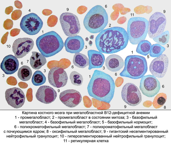
At B12-deficiency anemia in sternal punctate revealed a sharp irritation of red sprout, found large cells like red, and granular sprouts. Red cell nuclear peculiar shape, with the core, resembling minced meat, characterized by the difference in timing of maturation of the nucleus and cytoplasm. These cells are called megaloblasts.

The predominance of the sternal punctate basophilic and polychromatic megaloblasts regarded as "blue" bone marrow, It is indicating a severe form of anemia, and the prevalence of oxyphilic megaloblasts - as "red" bone marrow, indicating the onset of the disease or to make it easier to form. It was previously thought, megaloblasts that appearance - a sign of a return to embryonic hematopoiesis. More recently, it has been proved by modern research methods of radionuclide, megaloblasts that if B12-deficiency anemia are not special, shunt cell population, since in the presence of methylcobalamin they turn for a few hours at ordinary erythrokaryocytes. It means, that a single injection of cyanocobalamin or its derivatives can reverse the morphological picture of the bone marrow. Sternal puncture in this case does not allow a correct diagnosis, appear in the peripheral blood reticulocytes.
Diagnosis of cyanocobalamin deficiency
The largest errors in the diagnosis of vitamin B12-deficiency anemia due to the fact, that before the test at a hematologist or physician patient receives one or more injections of cyanocobalamin (derivatives). The examination of these patients revealed trehrostkovaya cytopenia, splenomegaly, hyperbilirubinemia by increasing indirect fractions and high content of reticulocytes, due to the introduction of cyanocobalamin (derivatives). Hematologist often sees these symptoms as manifestations of hemolytic anemia. When sternal puncture in this case revealed a sharp irritation and lack of red sprout megaloblasts. Direct Coombs test in this case is negative, but more sensitive machine-haemagglutination reaction may be positive, since it not only positive with autoimmune hemolytic anemia, and if B12-deficiency anemia.
All of these features are the basis for an erroneous diagnosis in patients with autoimmune hemolytic anemia, or its combination with autoimmune thrombocytopenia- or neutropenia. Often, these patients begin to heal prednisolone, giving at B12-deficiency anemia small temporary effect, It causes a decrease in the collapse of the nucleated cells of the bone marrow. In this regard, it is increasing the number of reticulocytes, reduced spleen.
Some time after the abolition of prednisone a recurrence of the disease. Patients, which have already been diagnosed with autoimmune hemolytic anemia, relapse is regarded as an exacerbation of autoimmune hemolysis, which entails the question of splenectomy, which is effective in exacerbations of autoimmune hemolytic anemia and absolutely not shown in the B12-deficiency anemia. Very often misdiagnosed autoimmune hemolytic anemia is put young patients, because it is considered to be, that B12-deficiency anemia occurs predominantly in elderly and senile. Really, aged 60 70 years, it has been an average of one for 100-200 people, in children aged from one year to 10 years - one on 10000, in adults aged 30-40 years - one on 5000. However, the recent trend towards the emergence of B12-deficiency anemia in young age. Such patients are particularly difficult to make a correct diagnosis.
Purpose of treatment cyanocobalamin (its derivatives) without special hematological study is a gross mistake. No less a blunder - this appointment cobalamin deficiency, folic acid, which, as indicated above, can lead to the development of funicular myelosis. Besides, under the influence of folate may temporarily disappear megaloblasts marrow, increase the level of reticulocytes, with the result that it is impossible to make a correct diagnosis. It should be remembered, Folic acid is a different multivitamin preparations. Hence, patients with unexplained anemia assign character are not to be. In the same extent this also applies to the appointment of raw liver.
Diagnose cobalamin deficiency in the body allow the microbiological methods for the determination of its content in the blood serum. The growth of a number of microorganisms Escerichia coli, Euglena gracilis, Lactobacilus leischmanis depends on the amount of serum methylcobalamin. Besides, for its definition used radioimmunoassay methods of investigation. Normal serum content of methylcobalamin - 200-1000 pg / ml. When vitamin deficiency level of the serum is reduced to 10-150 pg / ml.
To determine the absorption of cobalamin used radionuclide methods of investigation. Used cyanocobalamin, labeled with radioactive cobalt. The degree of absorption is determined either by the radioactivity of feces after taking vitamin, or by incorporation of labeled vitamin A in the liver, or on account of the total radioactivity of the body.
It is widely used Schilling method, comprising the steps of, that the patient give the labeled vitamin, and then injected into the body 1000 micrograms nonradioactive vitamin. Normally more 10 % administration of radioactive vitamin should stand in the urine. Malabsorption of vitamin urine radioactivity can not be observed. In this case, think of the violation or the secretion of intrinsic factor, or intestinal absorption of vitamin. In case of violation of secretion of intrinsic factor in the introduction of his body with the vitamin in the form of a concentrate leads to the normalization of vitamin intake, in violation of the intestinal absorption of intrinsic factor concentrate intake does not give effect.
For the diagnosis of cobalamin deficiency in the body is used method for determining urine methylmalonic acid. The study recommended after taking valine.
In all cases, cobalamin deficiency should be excluded invasion broad tapeworm. The stool is sometimes identified with the scraps strobill, found parasite eggs.
