Flatworms – description, morphology and development cycle
All representatives class of tapeworms, or cestode, look like a ribbon. Their body (strobil) consists of segments (proglotid), heads (scolex) and cervix. The causative agents of cestodiasis are divided into two groups: tapeworms and tapeworms.
Body length of different parasites, as well as the number of members (from three to four to several thousand), It varies between 0,5 mm to 10 and even before 20 m.
Head of helminths equipped with suction cups (fixation organs), in a number of tapeworms - chitinous hooks, in the tapeworm - with special suction slits - bothria. Digestive, respiratory and circulatory systems are absent. All cestodes are hermaphrodites: young penises are asexual, then they have male ones, and then the female genitals. As the individual develops, the male genital organs atrophy, and women's are rapidly developing. The mature penis has a uterus, filled with a huge number of eggs. The presence of a uterus is a differential feature - individual types of cestodes are recognized by its specific shape:
- in tapeworms, the uterus looks like a highly convoluted tube, opening on the surface of the joint;
- in tapeworms it is closed, saccular.
Eggs of tapeworms and tapeworms different structures: in tapeworms they have a cap at the poles and a thickening of the shell in the form of a tubercle, contain yolk cells along with germ cells, tapeworms have a delicate outer shell, sometimes it is equipped with special processes - filaments. Inside the egg there is a formed embryo (oncosphere) with its own rim and three pairs of embryonic hooks.
Formed tapeworm eggs due to the absence of an excretory opening, the uterus cannot freely exit into the external environment, and stand out together with the mature segments separated from the body (with feces). The eggs of tapeworms are freely released into the external environment, where the whole process of turning an egg into an embryo takes place. Most cestodes are biohelminths, develop with double (split) or triple (tapeworms) change of owners.
Wide tapeworm – description
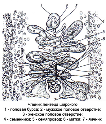
Wide tapeworm (Diphyllobothrium broad) - one of the largest human parasites, reaching in length 10 m or more. Head (scolex) its oblong-oval shape, flattened from the sides, has two deep slits - bothria, with which the tapeworm attaches itself to the intestinal wall. The body consists of many short and wide (at the head end) members, taking on a square shape towards the tail of the body, where they are more mature. In the center of the mature segments, the uterus filled with eggs is visible in the form of a rosette., having an outlet.
One adult tapeworm produces several million large tapeworms every day., oval gray eggs, sometimes yellowish.

The shell of the eggs is thin, smooth, double-circuit, with a cap on one pole and a small tubercle on the other. The egg is filled with many yolk coarse cells. It should be taken into account, that the eggs of the broad tapeworm are similar to the eggs of Nanophietus, however, in the latter they are more elongated, their shell is thicker and rougher, the cap is rougher, the tubercle is larger and rougher, protrudes very slightly above the surface of the shell.
Development cycle of the tapeworm occurs with a triple change of hosts. The final host may be humans, dog, cat, pig, bear, fox, seal, seal, walrus and some other animals. Intermediate host: freshwater crustaceans (cyclops), additional – freshwater fish (pike, we found, perch, ruff, etc.).
Broad tapeworm eggs are released into the environment with feces and must enter a body of water for further development, where the free-swimming larva matures in them (Coracidium) circular shape, with eyelashes. Coracidium is ingested by freshwater crustaceans, and those, in turn, fish.
In the body of fish, the larvae are carried to various organs and turn into a second form - plerocercoid; there are especially many such larvae in fish eggs. Plerocercoid is a small white worm up to 10 mm, having a head with bothria; he lacks a capsule and cilia; the body is not yet divided into segments. When eating insufficiently heat-treated fish or caviar, the larvae enter the human intestines, where within two months they transform into mature parasites.
Disease, caused by broad tapeworm, referred to difilloʙotrioz and usually occurs with an unclear clinical picture. Some patients develop pernicious-like anemia.
The diagnosis is made based on the detection of tapeworm eggs in the stool.
Bull tapeworm – Tapeworm unarmed – description
Bull tapeworm, or unarmed (Taeniarhynchus saginatus), reaches a length of 4-10 m. The round head has four suction cups; short, thin neck (tapeworm growth zone) goes into the body, divided into many segments. Younger segments are square, hermaphroditic, terminal mature - elongated, come off the strobile, mobile and can crawl out of the anus. The uterus has 18-30 thin lateral branches on each side of the median trunk and is completely filled with eggs (more 150 000).

Despite the daily separation of six to eight or more mature segments, the length of the tapeworm does not decrease, since more and more new segments are formed in the growth zone.
Bull tapeworm eggs round or oval, their shell is thin, transparent, Colorless, equipped with two thread-like appendages. There is an embryo inside the egg (oncosphere), equipped with three pairs of hooks and surrounded by a thick double-circuit, transversely striated yellowish-brown shell. The shell of released eggs is destroyed very quickly, therefore, during microscopic examination, only oncospheres are often visible, sometimes with remnants of a torn, wrinkled egg shell.
The final host of the bovine tapeworm is human, intermediate - cattle. Sexually mature individuals parasitize the small intestine, and the larvae are located in the intermuscular connective tissue. Due to the structural features of the uterus, without outlet, bovine tapeworm eggs are released into the external environment with the lower mature segments of the parasite, not only with feces during defecation, but also with active crawling of segments from the intestines. Eggs, released due to the destruction of joints, contain an already formed larva - oncosphere.
When the segments enter the external environment, soil contamination occurs, herbs, hay, livestock areas, pastures. Together with contaminated feed, oncospheres enter the food channel of cattle., where they are released from the shell, are introduced with the help of hooks into the capillaries of the intestinal wall, from where they can be carried with the bloodstream to any organs. The bulk of them settles in the muscles and turns into cysticerci (Finns) - white bubble-like formations up to 5 mm, filled with clear liquid. A scolex screwed inside is attached to the wall of the cysticercus on a thin neck. (appears as a white dot). Cysticerk enclosed in a capsule or cyst.
Finns can be stored in cattle muscles for one to two years, and then they die. A person becomes infected by eating raw or undercooked beef., containing Finns. In the human small intestine, the head of the finna turns out, attaches to the intestinal wall and after 2.5-3 months turns into an adult sexually mature parasite.
Disease, caused by an unarmed tapeworm - teniarinhoz - characterized by a variety of clinical manifestations, but sometimes the only sign of the disease is the release of joints during bowel movements (It is recommended to show the examinee preparations of the joints). Besides, macroscopic examination of stool is also carried out (identification of segments).
Teniarinhoz is diagnosed also by rectal or perianal scraping, since when the segments actively crawl out, the eggs are squeezed out, remaining on the walls of the rectum and on the skin around the anus. Conventional scatological research methods for teniarynchosis are ineffective, since eggs and oncospheres are rarely found in feces.
Pork tapeworm – Armed tapeworm – Description
Pork, or armed tapeworm (Blindfold seat) causes a disease similar to teniarhynchosis - tenioz.
Tapeworm length up to 3 m, the head is equipped with four suction cups and a double crown of hooks (29—32 small and large), neck thin, length up to 10 mm. Young penises are square, more mature - oblong. The uterus has 7-12 lateral branches, in mature segments it is filled with eggs, like the bull tapeworm.

In the eggs there is a formed larva (oncosphere) round shape, with fat, radially striated shell, inside which the embryonic hooks are visible. When examining the stool of patients with taeniasis, oncospheres are found (The delicate shell of the egg breaks easily).
An adult pork tapeworm parasitizes the human small intestine.. Mature segments separated from the strobilus are passively excreted with feces..
The transformation of the oncosphere into a cysticercus occurs in the body of the intermediate host (pigs, dogs, cats, wild boar, perhaps, and man). Formed in 2— 2,5 months cysticerci parasitize in various organs, but most often - in the intermuscular connective tissue. Man (final owner) becomes infected by eating raw or semi-raw meat from pigs, less common than wild boars, infected with cysticerci (Finns).
Diagnosis is based on the detection of diseased segments of pork tapeworm in the feces. Oncospheres of bovine and pork tapeworms are indistinguishable, therefore, when they are detected in feces in a laboratory report, they indicate the presence of taeniid eggs. A certain differential diagnostic sign is the absence of active crawling of segments.
Pork tapeworm can parasitize humans and in the larval stage, calling cysticercosis.
Oncospheres, in the stomach, freed from the shell, are introduced into the wall of the small intestine and are carried throughout the body through the bloodstream, settling in various organs and tissues, where they turn into cysticerci. They are most often found in the brain and eyeball, and in single copies, and thousands.
Cysticercus shape varies depending on location: in the muscles they are elongated, in loose fibrous connective tissue - rounded, in the brain they are flattened and have branches.
Methods of diagnosis diseases depend on the localization of the process: if the eyeballs are affected, ophthalmoscopy is performed; skin, subcutaneous tissue, muscle tissue - biopsy, calcified cysticerci are detected during x-ray examination. Cerebrospinal fluid examination and complement fixation test are also used..
Dwarf tapeworm – description
Dwarf tapeworm (Hymenolepis nana) reaches a length of 30-50 mm (most often 15-20 mm). Its body is whitish-grayish in color, tender, easily torn and consists of many small segments.

Mature end segments are filled with eggs, which, when the segments are destroyed, end up in feces in the intestines.
Eggs ellipsoidal shape, clear, Colorless, have thin, double circuit shell. Inside the egg is a transparent, colorless oncosphere, having a thin shell and three pairs of hooks. From the poles of the oncosphere thin, threads that sharply refract light - filaments.
Infection occurs through contaminated household items (pots, chairs, door handles, toys), when communicating with a patient. Once in the human digestive canal, the oncosphere is released from the egg and actively penetrates the villus, where it develops into a spherical larva - cysticercoid. Under the influence of waste products of the cysticercoid, the villi become necrotic, are destroyed or torn away from the mucous membrane and digested. The released parasites enter the intestinal lumen, where they attach to the mucous membrane and develop into adults (the development cycle lasts from 6-8 to 15 days).
The dwarf tapeworm is parasitic, usually, in children in the small intestine, rare in adults. There are cases of intraintestinal infection.
Diagnosis based on the detection of dwarf tapeworm eggs in feces. Due to the rapid destruction of eggs, fresh stool needs to be examined (no later than 3 h after isolation).
Rat tapeworm – description
Rat tapeworm (Hymenolepis diminutive) reaches a length of 0.2–0.6 m.
Eggs big, round or slightly oblong, clear, yellow color. Oncosphere is small, has six hooks and its own sheath. Definitive host: rats, mice and other rodents (a person can become the final owner by accident). Intermediate host - beetles, butterflies, fleas, millipedes.
A person becomes infected accidentally by ingesting grain and flour pests along with unbaked bread or other flour products., infected with tapeworm larvae. In humans, the rat tapeworm is a parasite in the intestines.
The diagnosis is made based on the detection of rat tapeworm eggs in the feces.
pumpkin tapeworm – description
pumpkin tapeworm (Canine dipylidium) reaches in length 0,7 m. Mature segments of the parasite resemble the shape of cucumber or pumpkin seeds.
Eggs very similar to dwarf tapeworm eggs, but differ from them in that, that several pieces are allocated (8-20) in one common shell - cocoon. This type of tapeworm usually infects dogs.. A person becomes infected by accidentally ingesting dog flea larvae., which are the intermediate host. In humans, the helminth parasitizes in the small intestine, causing dipylidia.
Diagnosis is made by microscopic examination of stool.
Echinococcus – description
Echinococcus (Echinococcus) - a very small parasite up to 2-6 mm long. His body consists of three- four segments, of which the first two are asexual, the third is a hermaphrodite, and the last one (the biggest) - mature and contains up to 800 eggs (according to other sources, up to 3000-4000). Helminth head has four suckers and 36-40 hooks, arranged in two rows.
The definitive host of the parasite is the dog., wolf, hyena, jackal, fox.
Echinococcus eggs or segments excreted in the feces of these animals (segments can crawl out of the anus and actively move around the host’s body, on the ground, grass) and pollute the wool, water, soil, grass. When eggs enter the small intestine of the intermediate host with water and food (most often mammalian or human herbivores) the larvae in them (oncosphere) develop to the invasive stage, are freed from the membrane and, with the help of hooks, penetrate into the blood vessels. They are carried by the bloodstream to the liver, where most of them linger, and then through the pulmonary circulation they penetrate into the lungs.
Eggs, caught in a big circle of blood circulation, can be entered into any body (most often in the brain and long bones). Settling in one or another organ, oncospheres form in fins, representing an echinococcal bladder, filled with fluid and containing a mass of tiny scolex.
Disease, caused by echinococcus in the finnosis stage, called echinococcosis.
The first three to five months the echinococcus bubble grows quickly, then its growth slows down and can last for years (10-20 years). Distinguish unilocular and multilocular echinococcus. Single-chamber echinococcus is a bubble the size of a millet grain to a child's head. Smaller daughter bubbles form within the bubble, containing a large number of heads. New bubbles can develop from daughter bubbles.
Multi-chamber cavity, or alveolar echinococcus filled with a conglomerate of small compressed bubbles with a gelatinous brown mass. The man is rare. The development of echinococcus in the intestines of the final host lasts 64-97 days, The lifespan of a parasite ranges from 6 months before 1 year and more. Larval forms of helminth (echinococcal blisters) retain viability in the body of intermediate hosts, including humans, for several (sometimes many) years.
Clinical echinococcosis caused by the pressure of the bladder on surrounding tissues and dysfunction of internal organs, toxic effects and sensitization of the body. If the echinococcal bladder ruptures, allergies may develop, including anaphylactic shock..
Diagnosis echinococcosis is based on the detection of mature segments or eggs of the parasite in the animal’s feces (dogs), suspected of being infected with echinococcosis. Diagnosis of echinococcosis in humans is very difficult, requires a comprehensive examination of the patient, including clinical observation, X-ray and laboratory examination.
Methods for diagnosing echinococcosis
Laboratory research methods include a general blood test, intradermal allergy test Kasoni, serological reactions (hemagglutination, latex agglutination, scolexol precipitations).
In a number of patients with echinococcosis, an increase in the number of eosinophilic granulocytes and ESR is observed in the blood.
Proba Kasoni quite specific, but sometimes, especially when repeated, may cause anaphylactic shock, which limits its use. Latex agglutination reaction is accessible and highly specific.
Echinococcal bladder punctate - transparent liquid of white or yellow color, in case of suppuration, leukocytes are found in it. Microscopic examination of the sediment after centrifugation of the punctate can reveal fragments of the shell in the form of grayish-white filmy formations, hooks and scolex.

When opening echinococcal blisters, depending on the location of the parasite in the feces, the contents of the duodenum, mokrote, urine can detect the same elements of echinococcus, less often - whole small daughter bubbles.
Alveococcus – description
Alveococcus (Echinococcus multilocularis) causes alveococcosis.
Its structure is very similar to Echinococcus, but differs from him in body size (1,3—2.2 mm), fewer and smaller hooks on the head. Alveococcus larva consists of a large number of microscopic bubbles, closely welded into one tight knot, That's why the parasite was called alveolar, or multi-chamber. The bubbles are filled with a viscous yellowish liquid or a thick dark mass. In animals, scolex are present in almost every vesicle., in humans they are less common.
The definitive host of the parasite is foxes, sand, less often - wolves and dogs. Intermediate hosts are rodents, sometimes a person.
Alveococcus development cycle almost the same, like echinococcus. Predators become infected, eating rodents, And they, in turn, become infected, ingesting alveococcal oncospheres, excreted in the feces of the definitive host. People can get infected from dogs, and also upon contact with wolf skins, foxes, pessov. Besides, infection is possible by eating berries, vegetables, water from natural reservoirs, where excrement from infected animals may end up (it is known, that alveococcal oncospheres are very stable in the external environment).
Clinical picture of alveococcosis characterized by the development of a tuberous liver tumor. The tumor grows into lymphatic and blood vessels, metastasizes to various organs and causes toxic-allergic reactions, possible development of secondary infection of a parasitic tumor.
Diagnosis alveokokkoz placed on the basis of epidemiological data, clinical observation, results of biochemical studies, immunological reactions, etc..