Characteristics of vaginal smears during physiological pregnancy
Before reading we encourage you to read: "The criteria for assessing the state of the vagina"
In the study of vaginal smears during physiological pregnancy found the same epithelial cells, as well as in non-pregnant women, t. it is. surface, intermediate and parabasal upper and lower layers. However, in different periods of pregnancy cytological characterization of vaginal smears is not uniform and depends on the degree of hormonal influences.
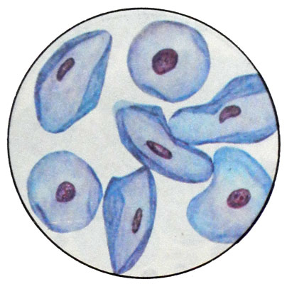
During pregnancy, estrogen production increases sharply, but, Despite this, in the vaginal smear dominated interim, instead of superficial cells. Besides, for pregnancy characterized navicular (boat-shaped) ovoid or boat-shaped cells with vesicular form, eccentrically located nucleus, which may have an elongated shape and being able pyknosis. These cells originate from the intermediate layers of the vaginal epithelium and are characterized by basophilic cytoplasm.

The predominance in smears of pregnant women due to epithelial cells of the intermediate high progesterone, continuing up to the prenatal period. Cells are intermediate groups and layers.
Since each gestational age corresponds to a certain kolpotsitogramma, it should be specified when the direction of the material on cytology. Without this interpretation of the results of research can not be colpocytologic.
In the early stages of pregnancy (5- 6 weeks) kolpotsitogramma corresponds to the luteal phase of the cycle. The smear is dominated intermediate cells, number of cell surface does not exceed 30 %. IS 0/70/30, CI and EI does not exceed 20-15 %. Cells are arranged in groups and layers. White blood cells and a small amount of sticks Dederleyna. Due to the lack of early pregnancy kolpotsitogramme specific changes in its use for diagnostic purposes at this stage is inexpedient.
As your pregnancy progresses content navicular smear and intermediate cells increases, The number of superficial cells, as well as CI and EI decreases. On the 8-10 th week of pregnancy the number of surface cells is not more than 20 15 %, IS - 0/80/20, CI and EI - 10 %.
Later in kolpotsitogramme marked change, which could be described by type of smears.
Strokes such as ongoing pregnancy
Strokes such as ongoing pregnancy appear after 14-15 weeks of gestation. They dominated navicular and intermediate cells, surface - in a small amount (5-7%); IS 0/93/7, EI to 1 %, THAT iniquity 3 %, but may be lower, up to 0. Parabasal cells are absent. Border cells in smears clear, basophilic cytoplasm. Leukocytes and sticks Dederleyna absent or present in small amounts. Swabs of this type are observed to 38-39 weeks of gestation. This is followed by prenatal hormonal reorganization: a reduction in the action of progesterone vaginal mucosa and the growing influence of estrogen, which causes corresponding changes vaginal smears.
Smears prenatal period
Smears prenatal period conventionally divided into three types:
- the approaching date of delivery;
- date of delivery;
- undoubted date of delivery.
Common to these types of strokes is loosening and loss of cell layers, change in color of the cytoplasm of a clear contrast to the pale.
Strokes such as approaching date of delivery
Strokes such as approaching date of delivery appear for 8-4 days before delivery. In them there is the same number of navicular and intermediate cells. The number of surface cells is 10-15 %, IS 0/85/15, EI - 5 %, THAT -6-10%. Detect single leukocytes and a small amount of mucus.
Strokes such as date of delivery
Strokes such as date of delivery can be observed for 3 days before delivery. In these intermediate cells prevail in relation to the navicular. The number of cell surface increases and reaches 25-35 %, IS - 0/65/35, EI - 8-10 %, THAT - e 20 %. The content of the mucus and leukocytes significantly increased.
Smears type undoubted date of delivery
Smears type undoubted date of delivery observed for 1--2 days before the birth and the day of birth. They are dominated by surface cells, and navicular missing. EI exceeds 20 %, THAT - 20-40 %. Strokes have the form of "dirty". Furthermore mucus and leukocytes occur in them erythrocytes.
Generally kolpotsitogramma prenatal (Pregnancy more than 36-38 weeks) characterized by certain qualitative changes in vaginal smears, although 15 % Pregnant change of the proportion of intermediate and superficial cells is not observed. However, the cell sheets have become looser, cells are often arranged in the form of rosettes, fuzzy outlines of cells. There are mucus and leukocytes. In some cases the color of the cytoplasm of intermediate cells become eosinophilic.
As with physiologically, and at the pathologically proceeding pregnancy can be observed cytolytic smears and inflammatory types. Morphologically, they correspond to the same types of strokes, as in non-pregnant women, and do not reflect the body's hormonal saturation.
Kolpotsitogrammu pregnant women should be investigated in dynamics. The need for re-examination is determined individually in each case.
