The structure and function of respiratory
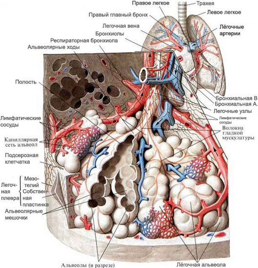
The respiratory system consists of the airways and respiratory (breathing) Department. By breather paths include the nasal cavity, larynx, trachea and bronchi. Respiratory Division presented acini, which includes respiratory bronchioles, alveolar ducts, ending in air sacs, and alveoli.

In the air-conducting ways is cleansed, humidifying and warming air, as well as to the regulation of the volume of air inhaled.
In the nasal cavity and respiratory distinguish eve region. The vestibule of the nasal cavity is lined with stratified squamous epithelium, which is a continuation of the skin epithelium. Under the epithelium in the connective layer are the sebaceous glands and hair roots of the nose. Respiratory region of the nasal cavity is covered with a mucous membrane, consisting of ciliated pseudostratified (multi-row ciliated) columnar epithelium and connective own records. The epithelium is composed of ciliated, intercalated epithelial cells and goblet cells.
Ciliated epithelial cells have cilia, represent cytoplasmic outgrowths height of about 3-5 microns.
Intercalary epithelial located between the ciliated, are on top of each other anastomosing microvilli size of 1.5-1.8 microns.
Goblet cells are unicellular mucous glands, allocating the secret to the surface of ciliated epithelium.
The mucous membrane of the larynx (except for the vocal cords), the trachea and the large bronchi is covered with pseudostratified cylindrical (prismatic) ciliated epithelium with a significant number of goblet cells. The bronchi medium caliber lined lower cylindrical pseudostratified ciliated epithelium with a small amount of goblet cells. In smaller bronchi pseudostratified (multirowed) ciliated epithelium is gradually becoming a two-lane and terminal bronchioles - single layer (single-row) cubic mucociliary. The respiratory bronchioles cubic cells lose their cilia. In a branching point trachea, bronchi and bronchioles pseudostratified ciliated epithelium is replaced by stratified squamous.
The bronchial mucosa are intercellular gap, which can detect cells, neutrophilic granulocytes, tissue basophils. The submucosa of the trachea to the bronchioles are mucous glands, particularly numerous in the bronchi medium caliber.
Respiratory bronchioles, branching, form alveolar ducts, each of which ends with two air sacs, consisting of the alveoli. This department is called respiratory respiratory. The wall of the alveoli, through which the exchange of gases between the external and internal environment (the air-blood barrier), formed, one side, capillary endothelial, on the other - the alveolar epithelium. The endothelium and epithelium are located each in their own basement membrane, between which there are elastic and collagen fibers, individual myofibrils, cells of the connective tissue and blood, including bone marrow-derived macrophages.
Epithelial, lining the cavity of the alveoli, It contains two types of cells: breathing (alveolocytes type 1) and large alveolar cells (alveolocytes type 2). On the free surface, facing the cavity alveoli, respiratory alveolocytes have cytoplasmic outgrowths, due to which the area of contact with air cells increases. In the cytoplasm of these cells are mitochondria and vesicles pinocytic.
Large alveolar cells have short cytoplasmic outgrowths. In the cytoplasm are larger mitochondria, Golgi complex (lamellar), osmophilic calf and the endoplasmic reticulum. They contain acid and alkaline phosphatase, nonspecific esterase and oxidative enzymes.
These cells have the ability to secrete into the lumen of alveolar lipoprotein substance (Surfactant), therefore they are also called alveolar secretory cells. From the walls of the alveoli, These phagocytic cells possess weak activity, However, it is possible, that the output of the lumen of the alveoli, it becomes more pronounced. In the lumen of the alveoli and alveolar phagocytes are (macrophages).
From the cavity alveolar surfactant alveolocytes covered - the thin film of surfactant, preventing alveolar spadenie output, and penetration through the alveolar wall of microorganisms from the inhaled air. Besides, surfactant prevents extravasation of fluid from the capillaries into the lumen mezhalveolyarnyh walls of the alveoli. The surfactant is distinguished membrane portion, composed of phospholipids and proteins, and liquid, representing glycoproteins.
Membrane phospholipids surfactant synthesized and secreted large alveolocytes into the lumen of the alveoli merokrinovomu type secretion, and alveolar phagocytes are involved in the removal of surfactant with respiratory surface.
The deficit in the alveoli of surfactant phospholipids may be accompanied by the development of atelectasis, the appearance of acute respiratory failure. Excessive accumulation of surfactants is accompanied by decrease of the surface tension in the alveoli, resulting in difficulty exhaling and increased residual lung volume, contributing to the development of emphysema.
Mucous glands and goblet cells of the bronchi a healthy person constantly produce the necessary amount of mucus, which is subjectively felt. It provides a physiological barrier between the inhaled air and alveolar cells, protecting them from the damaging effects of environmental factors. Increased mucus secretion in response to irritants of various origins (dust, smoke, gas, infection, etc.. d.) is protective in nature. Prolonged exposure to the irritant factor develops hyperplasia of mucous glands of the bronchi with the increase in their number of secreting cells. So, in the distal bronchi appear goblet cells, that they lack. Besides, and increases the intensity of the development of a secret. As a result of hypersecretion of mucus increases dramatically its number, which leads to a violation of the drainage function of bronchi, which under physiological conditions is provided mucociliary escalator action, reduction of bronchial and cough stimulus.
With an increase in the viscosity of secretions especially disturbed mucociliary escalator function. This is compounded by the appearance in the distal bronchi, where no physiological way of removing slime, goblet cells, producing particularly viscous secret. Violation of the drainage function of bronchi leads to the accumulation of mucus in the form of sputum.
