Structure and function of the spleen
Spleen covered serous membrane, under which there is fibrous sheath. From inside the fibrous sheath of the spleen depart crossbar (trabeculae). The space between is filled with white and trabeculae red pulp. The white pulp of the spleen It is a collection of lymphoid tissue, arranged around central arteries (Lymph follicles of the spleen).
Lymph follicles of the spleen distinguish periarterial zone, mantle layer and the edge, or marginal, area.
Periarterialynaya area It occupies a small portion of the follicle around the arterioles. In functional terms, it is analogous substance okolokorkovomu lymph nodes, and is composed of T-lymphocytes and cells interdigitiruyuschih, processes which are contacted with T-lymphocytes. Role interdigitiruyuschih cells is, they absorb the antigen and stimulate proliferation and blast transformation of T-lymphocytes.
Mantle layer It consists of a small T- and B-lymphocytes, which form a kind of crown, exfoliated circularly directed reticular fibers. It is believed, that this zone plays an important role in humoral immune responses.
Breeding Center follicle identical same lymph node follicles. It consists of proliferating B-lymphoblasts, reticular cells and macrophages. With a large number of macrophages central part looks bright follicles (Jet Center).
Marginal, or marginalynaya area - Is part of the transition between the white and red pulp. It consists mainly of T- and B-lymphocytes and macrophages single, who first met with antigens. B lymphocytes that zone after contact with macrophages transformed into plasma cells.
Billroth's cords It consists of the end of the capillaries, venous sinuses and are located between the reticular fibers, among which are free to the various cells. A large number of red blood cells attached to the red pulp. In the reticular fibers are constantly found with fragments of macrophages cells, grains or clumps, and hemosiderin microorganisms.
The sinuses of the spleen lined with rod-endothelial. The walls of the sinuses (at the transition into the vein) are muscle zhomy. When relaxing the arterial and venous blood flows freely zhomov veins, while reducing their blood shall be deposited in the sinuses. This is the destruction of red blood cells and moribund enrichment serum mineral and organic substances. Relaxation zhomov promotes exit of blood in venous bed. The outflow of venous blood from the pulp of the spleen occurs through the trabecular veins, representing a narrow gap, lined with endothelium, which facilitates the release of the blood while reducing spleen.
The spleen is a universal blood-forming organs in the embryonic stage of development, and produces lymphocytes after birth. With the function lymphopoiesis closely linked to develop the ability of spleen immune body. The protective function of the spleen is also carried out through its ability to phagocytose foreign particles and microorganisms. The spleen has lytic properties, and therefore fall within its obsolescent erythrocytes undergo hemolysis.
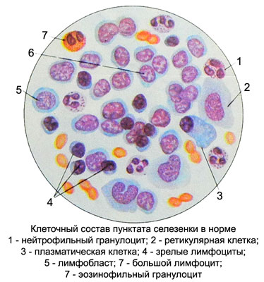
In the spleen, delayed blood and iron. Expected, that it produce substances, suppress bone marrow erythropoiesis. The cellular composition of the spleen punctate It varies widely even in the case, if the preparations are prepared from different portions of the same spleen.
Cytogram spleen punctate OK, %
- Lymphoblasts - 0.1-0.2
- Prolymphocytes and lymphocytes - 60-85
- Large reticular cells - 0-0.1
- Small reticular cells (not always reliably determined) - 1-10
- Plazmoblasty, proplazmotsity and mature plasma cells - 0.1-0.8
- Macrophages - 0,1-0.2
- Lipofagi - 0,1
- Monocytes - 1.5-2.5
- Rod-endothelial (pulp cells) - From 0.2 to 0.6
- Tissue basophils - 0-0.1
- Myelocytes and metamyelocytes - 0-0.1
- Neutrophil stab and segments- nuclear granulotsity- 10-15
- Eosinophilic granulocytes - 0.5-2
- Basophilic granulocytes - 0.1-1.5
Thus, among cellular elements predominate punctate spleen cells lymphoid series.

The bulk of them make mature lymphocytes, among which there are large shirokoplazmennye, reaching in diameter 17 m (their normal 6-8 %), a small number of lymphoblasts and prolymphocytes.
From cell reticulum should be noted large and small reticular. Large reticular cells found in the spleen punctate only in pathology (hemolytic anemia, myelogenous leukemia, etc.. d.). Small reticular cells They are a normal part of splenogrammy, but often difficult to distinguish from lymphocytes.
Plasma cells in the normal splenogramme rare. However, in certain diseases, especially in chronic inflammatory processes, their number is increasing, where you can find all the transitional forms.
Macrophages (pigmentofagi, lipofagi, bacteriophages) rarely found, tissue basophils - even rarer, but in chronic inflammatory processes their number increases.
Rod-endothelial (pulp cells) specific to the spleen. They are oval or elongated, sometimes tailed form, diameter 20-30 mm. The cell nuclei are compact, adelomorphous, in young cells contain one or two nucleoli; cytoplasm is colored gray with a purple tinge, often contain haemosiderin. Cell layers are arranged. They are closely linked, which facilitates the process of differentiation. When there are disease states and separately laying cells. In cytological preparations distinguished from reticular endothelial cells is quite difficult.
Serous cells. These include mezoteliotsity, covering serous membrane of the spleen. When spleen puncture mezoteliotsity can get into punctate. Usually mezoteliotsity have small clusters.
Blood cells. Normally, in addition to spleen punctate mature blood cells can be detected and isolated erythrokaryocytes myelocytes. In some blood diseases and disorders of the hematopoietic function of bone marrow (osteosclerosis, tumors of the bone marrow) spleen extramedullary hematopoiesis observed.
