Structure and function of the skin
Skin is the outer covering of the human body, and in addition to the protective performs a number of vital physiological functions - breathing, excretory, Temperature- Thorn, chromogenic, takes involved in metabolic processes and reflex reactions, providing tactile and thermal sensitivity, and etc.
The skin consists of epidermis and dermis (dermis), under which there is subcutaneous fat.
The epidermis of the skin
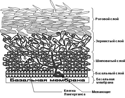
Epidermis Skin is represented by stratified squamous epithelium of the stratum, consisting of five layers of cells:
- Basal;
- Shypovatoho;
- Seedy;
- Brilliant;
- Corneous.
The basal layer of the skin It located directly on the basement membrane and consists of a single layer of basal cells, among which are the pigment cells - melanocytes. Basal cells are small sizes, cylindrical shape, in Pappenheim-stained preparations are painted sharply basophilic cytoplasm, oval or elongated nucleus with a lot of chromatin.
Melanotsitы sinteziruyut pigment - melanin. It is process cells with dark-colored core and the light basophilic cytoplasm, wherein grains are melanin.
Spiny layer of the skin It located above the basal and comprises 5-10 series spiny epidermotsitov. This polygonal cells with numerous cytoplasmic processes. The cells of the basal and spinous (Deep divisions) multiply layers of mitotic division, why they are united under the name sprout (embryonic) layer. The epidermis is updated every 19 20 days.
The granular layer of the skin located on the sprout. It consists of three or four rows of relatively flat cells, cytoplasmic granules which are keratohyalin. Keratohyalin It is a protein with a large number of basic amino acids (arginina, lysine, gistidina, cystine), It is formed from keratin - keratin.
Shiny skin layer It consists of three or four rows of flat cells, kernels are not colored. The cytoplasm of these cells impregnated with a protein substance - eleidin, formed from keratogielina and contain many disulfide groups, give the skin shine.
The stratum corneum of the skin consists of a plurality of rows of dead cells - horny scales. The flakes contain keratin - a dense substance, than eleidin, and air bubbles. Keratin - A protein with lots of sulfur, very resistant to various chemicals. The stratum corneum is completely renewed every 7 11 days. Describe the structure of the skin is the epidermis of the palms and soles, in others it is thinner. With a lack of retinol in the body (vitamin A) dramatically enhanced the processes of keratinization.
The structure of the dermis
In your own skin (dermis) distinguish the papillary and reticular layers, without a clear boundary between.
The papillary layer skin located immediately below the epidermis and consists of loose connective tissue unformed fiber, which forms numerous papillae, vdayuşçïesya in the epithelium. Most papillae in the skin of the palms and soles, in the face, they are very poorly developed and may disappear with age. The papillary layer determines the individual character of the pattern of the skin.
The connective tissue of the papillary layer It includes collagen, elastin and reticular fibers, connective tissue and reticular cells, gistiocitov, fibroblasts, macrophages, tissue fat cells and basophils, neischerchennyh muscle fibers and the pigmented cells of process (melanophore).
Net layer of the skin formed by bundles of collagen and elastic fibers, arranged in a network. Cells mesh layer consist principally small amounts of fibroblasts, near vessels lie macrophages and leukocytes. In most parts of the reticular layer of the skin are the sweat, sebaceous glands and the hair roots. Net layer determines the strength of the skin.
Bundles of collagen fibers a mesh layer penetrate into the subcutaneous fat. Since not only contain melanocytes, but also form a pigment, they are called melanoblast. These cells give positive DOPA-reaction. The proper skin pigment located in the cytoplasm of cells Process forms. They can not synthesize the pigment, do not give positive DOPA-reaction, therefore they are called melanophores (xromatoforami). Melanophores larger granules comprise pigment, than melanoblast, and are located only in the mammary areola and the anus. In albino pigment in this area offline.
The glands of human skin
In human skin are the sweat and sebaceous glands.
Sweat glands - Simple tubular, merokrinnye, part of apocrine, sebaceous - simple alveolar, The branched end portions, golokrinnye, nearly, always associated with hair.
Secretory processes of the skin provide thermoregulation. Fat sebaceous glands It protects the skin from drying and the harmful effects of various substances; glands of the skin secrete some products of metabolism (Urea, uric acid, ammonia and others.), and entering the body drugs and toxic substances.

