Preparation and microscopic examination of lymph nodes punctate
Puncture study lymph node It is available, technically easy to implement and allows you to observe the process in dynamics, Unlike histological examination, wherein lymphatic node is removed surgically (biopsy).
An important condition for the puncture of a lymph node is a reliable fixation of his fingers to the muscle or bone, as the pressure of the needle assembly is immersed deep. When puncture of large dense lymph node should use a thick needle with a wide clearance.
The syringe and needle should be dry. Before puncture alcohol or alcoholic solution of iodine disinfect skin, a fixed input unit needle and syringe plunger nasasyvayut movements needed to get the study material. After each movement of the syringe is removed nasasyvatelnogo. Sometimes the material is so small, he gets only the lumen of the needle. The contents of the syringe needle and movement of the piston is pushed onto the slide (If punctate meager) or in a petri dish. Syringe with needle washed in another Petri dish with isotonic sodium chloride solution to be detected in the wash water the smallest particles tissue, that can stay on the walls of the syringe, Piston or the needle.
Punctate study macroscopically and described, then collected tissue particles and from cooked preparations for microscopic examination. If punctate meager, he was immediately placed on a slide, covered with a cover and explore. First, learn the native drugs at a small and a large increase in, and then remove the cover glass, stretch material with a thin layer on a glass slide and stained.
If you find native preparation of pus is necessary to prepare smears for Gram stain and Ziehl-Nelsenu, If found detritus, his stained by Ziehl-Nelsenu. If the cyst lymph node, aktinomikoze, metastasis-differentiated squamous cell cancer diagnosis is based on the study of native products. In all cases, after a study of the native preparations prepare smears for painting. Their dried, fixed and stained by conventional methods and study initially under low magnification, and then using an immersion system.
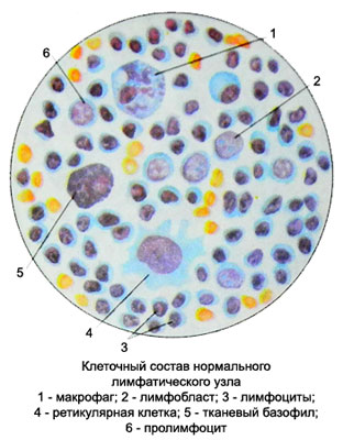
The punctate normal lymph node 95-98 % comprise lymphoid cells, most of them - mature lymphocytes, no different from blood lymphocytes.
Along with mature cells and blasts across individual prolymphocytes. The transition to a mature lymphocytes is accomplished through a stage of large lymphocytes. The punctate normal lymph node macrophages found, phagocytic damaged cells or particles, pigment, bacteria, fats and lipids, plasma cells, tissue basophils, sometimes eosinophilic granulocytes. Neutrophil granulocytes are found in punctate of lymph node in the event, if they fall back from the blood during the puncture.
To make out the results of the study can be in the form of punctate cytogram (in different parts of the stroke calculated 800-1000 cells and displays their percentage) or descriptively (tuberculosis, limfogranulematoze, sarcomas, metastatic cancer, and others.). In both cases, there is the number of punctate and macroscopic characteristics (bloody, saniopurulent, festering with small, grayish or grayish-whitish shreds, etc.. d.).
At the end of the description, or cytogram provide a conclusion on the nature of the pathological process.
Changes in the lymph node during pathological processes They do not always have the characteristic features. When an infectious process develops lymph node hyperplasia. Thus in a punctate increased number of mature and less mature lymphocytes (mainly large) and to a lesser extent prolymphocytes and lymphoblasts, and plasmatic, reticular and mast cells. With increasing number of hyperplasia less mature lymphocytes and reticular cells increases. With further development of the disease cellular structure becomes punctate characteristic for each disease.

