Nephrolithiasis – state and urinalysis
Nephrolithiasis - A relatively common disease (It is between 25 to 50 % all diseases of the urinary organs). In most cases, they suffer from age 25 to 40 years. In most cases stones are formed in one kidney, Bilateral renal involvement occurs in 15 % patients. Kidney disease usually affects men.
By the etiological factors of the disease should include infection, metabolic disorders, feeding, endocrine disorders, climatic and geographical features (iodine deficiency, limit water consumption, dry hot climate, and so on. P.).
At the heart of stone formation in the urinary tract is a violation of colloid-crystalloid balance. The urine retained in the salt dissolved due to the presence of protective colloids,. They represent a variety mukoproteidov - glycosaminoglycans (mukopolisaxaridы), or high molecular weight polysaccharides, containing protein components - aminosugar. With enough content in the urine of protective colloid crystal formation in supersaturated solution inhibited. Otherwise, the urine formed micelles - future core stones. They may be formed from fibrin, blood clot, cellular debris, bacteria, other components of urine and foreign bodies, located in the urine. The deposition of salts on the original nucleus depends on their concentration, the reaction of urine, the amount and qualitative composition of urine colloids. Kamen- Education takes place by gluing salt crystals cementing foundation, give the stone a stable structure.
When the acid reaction of urine observed stones, consisting of salts of uric acid, when alkaline - from phosphates; oxalates may occur both in acidic, and in the alkaline environment. Perhaps the formation of protein, cystine, Xanthinovs, sulfanilamide and other stones.
The appearance of stones in renal pelvis causes catarrhal changes in the mucous membrane. The process begins with hyperemia and edema of the mucous membrane of the renal pelvis. Then hemorrhages appear on it. The function of the renal pelvis and ureter is violated, What is expressed in pyeloctasia and the expansion of the ureter below the level of the location of the stone. As the process progresses, the kidney parenchyma is subjected to fibrous degeneration.
During the disease, there is an interconnectional period and renal colic.
Renal colic is accompanied by hematuria. During renal colic, rapid painful urination is observed. The allocation of urine is reduced, up to anuria.
With the non -infection of urine stones in the interconnectional period or normal, Or there is a microhematuria. After an attack of renal colic or during it in the urine, red blood cells and protein are found in a larger or smaller amount.
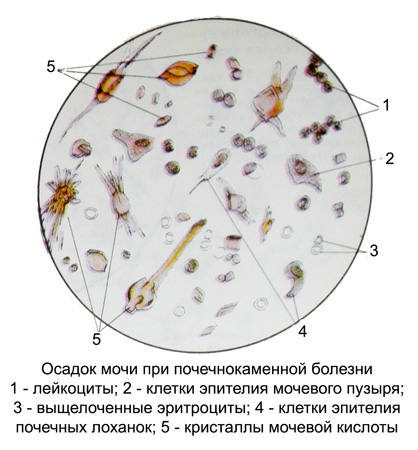
With a microscopic examination of urine sediment, a normal or slightly increased amount of leukocytes and red blood cells is detected.

The red blood cells are usually leaned out, and with macrohematuria - and unchanged. Fibrin brown -colored, Klochko. Cells of transitional epithelium of renal pelvis are found separately and groups (imbricate arrangement), sometimes quite large, and scraps. There may be hyaline and granular casts and single cells of renal epithelium. Salt crystals or form aggregates, or have a spear, bayonet and another form, with the sharp angles of crystals are the cause of hematuria. Sometimes found crystals gematoidina, lying freely or necrotic scraps. With the development of pyelonephritis appears urine, typical for this disease. When clogging the ureter with a stone in the renal pelvis, pus accumulates and pionofrosis develops.
In some cases, the disease proceeds asymptomatic. However, in the urine in such patients, especially after physical exertion, more or less red blood cells and salts are detected. With bilateral calculent pyelonephritis, the outcome of the disease may be the kidney failure.
