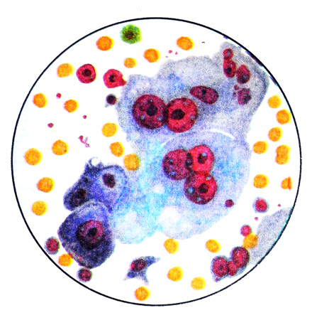
Kidney Tumors – state and urinalysis
Among the malignant renal tumors in adults, the most common cancer, histological structure in which you can select five options:
- clear cell;
- granular cell (dark cell) solid-tubulyarnыy;
- spindle cell (polymorphonuclear cell) sarcomatoid;
- glandular (the usual type of adenocarcinoma);
- mixed.
Kidney cancer observed in adults is relatively rare and is 1- 2 % All tumors. Pediatric renal tumors occur much more frequently (20-25 % cases). Men (mostly aged 40 to 60 years) ill almost twice as likely to, than women. The right and left kidneys are affected with equal frequency. Bilateral tumors are observed only rarely. Primary tumors of the kidney are more common, than metastatic.
The most common histological form of kidney cancer in adults is svetlokletochnыy cancer. In cytological preparations with the most frequently detected quite large polygonal cells with pale vacuolated mesh, sometimes like an empty cytoplasm.

Most of the cytoplasm occupied by the inclusion of glycogen and neutral lipids; cytoplasmic organelles are scarce. The content of glycogen and lipid in cancer cells is regarded as a manifestation of the peculiarities of their metabolism. The cell nuclei are usually small, large or oval, hyperchromatic, located centrally or more eccentrically.
Sometimes drugs are found small and medium-sized cell cubic, round or polygonal shape with a darker colored granular basophilic cytoplasm and a round or oval nucleus in the cytoplasm of these cells is a small amount of glycogen and lipid.
Against the background of the expressed cellular polymorphism can meet giant ugly symplast. It granular cell (temnokletochny) cancer.
Spindle (polymorphonuclear cell) sarkomopodobnыy cancer characterized by the presence of spindle or polymorphic cells with vesicular nuclei and compact or granular or vacuolated cytoplasm. Among the described cellular elements can meet the typical bright cells.
In mixed cancer there may be all kinds of cells described.
Pediatric renal tumors up 1/3 of all malignant tumors. Frequently (in 20 % cases) found Wilms tumor or nephroblastoma. It appears this tumor in children mainly in the first years of life, sometimes it can be congenital.
Neoplasm localized in different parts of the kidney, it usually has a dense fibrous capsule. Consistency - from dense to myagkoelasticheskoy. Expanding, the tumor compresses the renal parenchyma, gradually causing its complete atrophy. The surface of the tumor is often uneven. In the context of often visible areas of necrosis and softening to form cavities, hemorrhages, occasionally with areas of calcification and ossification. The tumor metastasizes very early, usually the lungs.
Histology Wilms tumor is typically characterized by the presence of tubules, lined by cubic or columnar epithelium, cells which have large hyperchromatic oval nuclei and narrow bezel basophilic cytoplasm. In some cases, there are rolls with a wide lumen and cyst, in other cases, or in other sections of the same tumor lumen tubes almost indiscernible. Tubular structures may not have the basal membrane and move into the broad field of solid or solid strands sprawl round, oval or spindle-shaped cells with a rather dark nuclei, as well as the structure rozetkopodobnye. Sometimes, there are pockets of proliferation of undifferentiated cells extended, fusiform with scant cytoplasm, many of which are in mitosis. These cells are often located beams, as in sarcoma. Hence the name of the tumor - adenosarcomas. A similar pattern is observed in tumor punctates.
The urine in the Wilms' tumor can get small cell limfotsitopodobnye, which is not easy to differentiate, since they are similar to leukocytes. Kernels take them almost the entire cell, nucleoli are viewed poorly, and basophilic cytoplasm has the form of a narrow, subtle rim. Clusters of cells are arranged in groups and close.
Sarcoma of the kidney
On histological structure in the kidney is most common fibrosarcoma varying degrees of differentiation. Elements fibrosarcoma rarely found in the urine and are a group of cells in soft semi-transparent scraps. In the study of stained preparations under the microscope with immersion objective fibroplastopodobnye revealed the same type of cells with varying degrees of malignancy.
Bacterioscopic urine when kidney sarcoma manufactured using staining Ziehl-Nelsenu to detect Mycobacterium tuberculosis and Gram - for identifying Escherichia coli and Neisseria gonorrhoeae.
