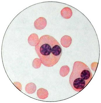
Hereditary anemia dizeritropoeticheskie, related to the violation of the fission process erythrokaryocytes
Hereditary anemia dizeritropoeticheskie - A group of rare forms of anemia, characterized by signs of violation of the division erythrokaryocytes, intramedullary destruction of their, plenty dual-core or multi-core erythrokaryocytes in the bone marrow, sharp stimulation of red bone marrow sprout at a relatively small reticulocytosis. There are three types of hereditary anemia dizeritropoeticheskoy:
Type I is characterized by megaloblastic bone marrow, dual red cells, internuclear chromatin bridges between sections of cells.
When II and III types of anemia dizeritropoeticheskoy megaloblasts in the bone marrow are missing. When you identify the type II dual-core and triple-core erythrokaryocytes, found karyorrhexis.
Type III is characterized by a pronounced macrocytosis, the presence of giant erythrokaryocytes, containing from 5 to 12 cores. The most common anemia type II. Currently, the literature describes a 75 patients with this form of anemia.
Dizeritropoeticheskaya anemia type I and II inherited in an autosomal recessive, and type III - likely an autosomal dominant, Currently, she is described only in a few families.
The etiology and pathogenesis of anemia dizeritropoeticheskoy until specified. Expected, that the cause of violations of nuclear fission is a defect mechanism, underlying the termination of nuclear fission under the influence nor gemoglobinizatsii- motsitov.
The clinical manifestations of anemia dizeritropoeticheskoy
Patients dizeritropoeticheskoy anemia often sent to the hospital with a diagnosis of "chronic hepatitis" or "hereditary hemolytic anemia". There yellowness of varying severity. There are changes in the skeleton - high-top sky, Skull Tower, a short little finger.
In most patients, increased spleen, sometimes significantly.
Laboratory tests at dizeritropoeticheskoy anemia
When I type dizeritropoeticheskoy anemia hemoglobin is 4,96-7,45 mmol / l (80- 120 g / l), most patients anemia type II 4,96-6,21 mmol / l (80- 100 g / l), however there may be cases decline to 3,1 mmol / l (50 g / l). When you type III dizeritropoeticheskoy anemia hemoglobin level above 5,59 mmol / l (90 g / l).
Anemia, usually, normohromnaja. The content of erythrocytes in 2-3.5 G 1 l. The number of reticulocytes increased slightly (to 2.5-3.5%) or within the upper limit of normal (1,2-1.8 %). When I and III types of anemia dizeritropoeticheskoy marked macrocytosis, pronounced anisocytosis, fragmented erythrocytes.
With type II disease often Anisocytosis, sometimes moderate hypochromia, basophilic erythrocyte punktatsiya, occasionally microspherocytosis. The content of normal leukocytes and platelets. Leukogram without deviation from the norm.
In the bone marrow there is a sharp erythroid hyperplasia. Leykoeritroidnoe ratio often less than one. When all three types of the disease in most patients erythroblasts morphologically different from normal. Changes begin in basophil normocytes. When different types of anemia dizeritropoeticheskoy they are diverse.
When I type about anemia 10 % polychromatic and oxyphilic normocytes have dual core. There are also multi-normocytes. Some fragmented nuclei, part with signs karyorrhexis. Characteristic of chromatin bridges between the nuclei section norm- tsytamy. For the type I deficiency anemia tend to morphological similarity with erythrokaryocytes megaloblasts. When painting on iron revealed a large amount of it in macrophages, normocytes in iron content also increased, but the location of its ring around the core is not typical for this disease.
For dizeritropoeticheskoy anemia type II are not typical megaloblasts. Part of the polychromatic and oxyphilic normocytes contains two cores, Some 3.4 kernel.

Among basophilic normocytes can also be dual-core. Dual erythroblasts not found. In oxyphilic normocytes found karyorrhexis and lobed structure of the nucleus, there are cells with nuclei, napomynayuschymy tutovuyu berry. In some cases dizeritropoeticheskoy anemia type II shows a sharp stimulation of red bone marrow germ with a small number of reticulocytes. This dual erythrokaryocytes are no more than 2-3 %.
A characteristic feature of dizeritropoeticheskoy anemia type III is the presence of giant erythrokaryocytes, with up to 12.5 cores.
Electron microscopy of bone marrow anemia type I and II revealed various anomalies cores erythrokaryocytes, clippings from mild to deep cracks. In some cells the nuclear envelope loses its alkalinity and the cytoplasm into the nucleus between chromatin masses. A characteristic feature of dizeritropoeticheskoy anemia type II is the presence of a double membrane in the cell. In some cases, additional membrane forms a kind of oval inclusions, lying in the region of 40-60 nm from the inner surface.
On the surface of red blood cells of patients with anemia type II dizeritropoeticheskoy found an unusual antigen, against which serum many healthy individuals have antibodies. The presence of such antigen can be explained by changes in the structure of membrane proteins, identify them by electrophoresis gel polnakrilamidnom, especially when using two-dimensional electrophoresis. In the sera of healthy individuals contains certain natural antibodies hemolysin type, directed against the antigen, detected in patients with anemia type II dizeritropoeticheskoy.
Under the influence of red blood cells of patients with complement dizeritropoeticheskoy anemia type II are destroyed as well, as well as red blood cells of patients with paroxysmal nocturnal hemoglobinuria, but all APG odnogruppnoy fresh serum donors by having them complement destroying the red blood cells, and if the disease had devastating effects on red blood cells have only the serum, which contain antibodies against erythrocytes of patients with anemia dizeritropoeticheskoy.
Dizeritropoeticheskaya anemia type II often found in the literature as HEMPAS.
A characteristic feature of dizeritropoeticheskih anemia is an excess of iron in the body. The iron content increases sometimes up 54 mmol / l (300 g%). Some patients found moderate hepatic siderosis.
Radioactive iron, administering to a patient, quickly leaves the plasma, However, the inclusion of iron in the red cells with reduced (25-50 %). There radioactive iron accumulation in the bone marrow, indicating that the so-called ineffective erythropoiesis, t. it is. a pronounced medullary destruction erythrokaryocytes. This is also indicated expressed irritation red growth marrow with a small increase in the level of reticulocytes.
Bilirubin increased mainly due to indirect, and also due to bone marrow failure normocytes. This is also evidenced by the increasing level of carbon monoxide in exhaled air. The value of this parameter corresponds to the degree of destruction of hemoglobin.
