Microscopic examination of the bile
Microscopic examination reveals elements of inflammation, violation of colloidal stability of bile and parasitic infestation.
For the preparation of drugs bile poured into petri dishes, needle spatula and taken lumps of mucus, placed on glass slides, cover their cover glasses and examined under low and high magnification. In the absence of lumps drugs made from bile sediment after centrifugation.
In normal bile microscopic elements almost undetectable. If the pathologist finds lumps of mucus, cell, crystalline formations, parasites and bacteria.
Slime in the form of small lumps found in catarrh of the biliary tract and duodenitis.
White blood cells may fall into the contents of the duodenum from the mouth, Respiratory (with phlegm), stomach, gallbladder and biliary tract. Regardless of their place of origin when released into the bile they quickly painted under the influence of bile acids and soaps are destroyed.
The most common cause of duodenal contents of leukocytes is duodenit. In such cases they are often surrounded by a cylindrical ciliated epithelium cells of duodenal mucosa.
Diagnostic value of an inflammatory process in the gall bladder is only the presence of white blood cells, found in lumps of mucus in the portion B together with high prismatic ciliated epithelium of the gallbladder (mucus partially protects leukocytes from the destructive action of bile). If cholangitis in the mucus can identify leukocytes and epithelium of the biliary tract.
Eosinophilic granulocytes contents of the duodenum are found in allergic cholecystitis, cholangitis, and helminths. They are more resistant to the destructive action of bile, than leukocytes.
In addition to the items in the contents of the duodenum can detect exfoliated epithelial cells of the mucous membrane of the gallbladder and biliary tract, stomach, duodenal, oral, respiratory.
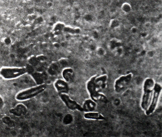
For cholecystitis characterized by the presence of prismatic ciliated epithelial cells, for cholangitis - small prismatic and liver epithelial cells resembling comma strokes or high prismatic epithelial cells of the common bile duct, spaced apart and lumpy mucus (often in combination with leukocytes). The discovery of large cylindrical epithelial cells with cuticle indicates a pathological process in the duodenum.
The contents of the duodenum can be identified leykotsitopodobnye education - leykotsitoidy, which differ from the large size of leukocytes and negative reaction to peroxidase.
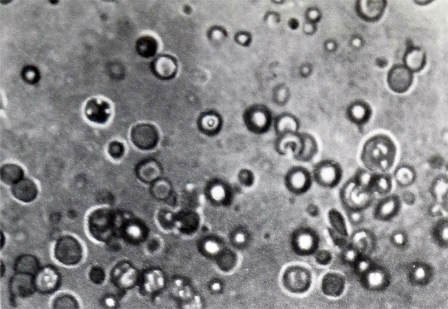
Suggest, they are modified cylindrical epithelial cells of the duodenum. Diagnostic value of not found. Do not have a diagnostic value revealed squamous cells and macrophages. Sometimes the contents of the duodenum can detect malignant abnormal cells, that allows you to diagnose malignant tumors of the duodenum, stomach, biliary tract and gall bladder.
Crystal formation in the bile
Cholesterol crystals found in normal bile rarely, in a small amount. The abundance of cholesterol crystals indicates a change in the colloidal stability of bile. Along with the other crystalline form crystals of cholesterol observed in the bile in cholelithiasis. Most often they are in the form of lumps.
Microlites It is a dark compact rounded education and multifaceted form, consisting of salts of calcium, mucus and cholesterol (for their identification, preparations of mucus and bile sludge).
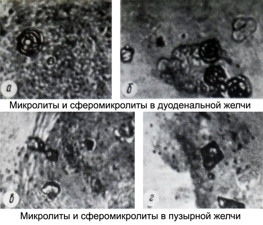
Normally microlites not found in bile. They can be found in cholelithiasis, often with cholesterol crystals, fatty acids and bilirubin calcium.
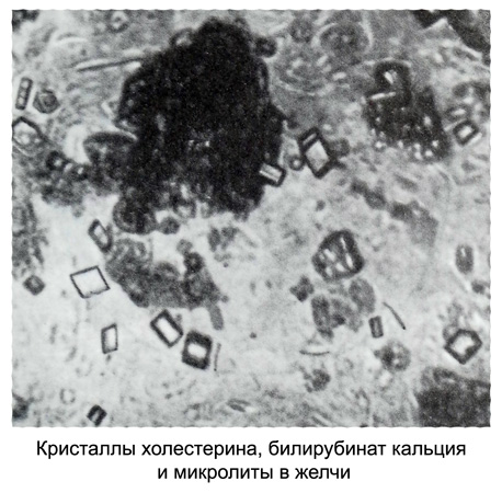
Fatty acids They have the form of needles, Sometimes clumps, often identified with cholesterol, microlites, soaps and calcium bilirubinate when changing the colloidal stability of bile and decrease the solubility of fatty acids by reducing the pH of bile yell inflammation in the gallbladder.
Calcium bilirubinate takes the form of small grains of golden-yellow and brownish, precipitate, sometimes visible macroscopically; detected in preparations, made from sediment of bile or bile lumps.
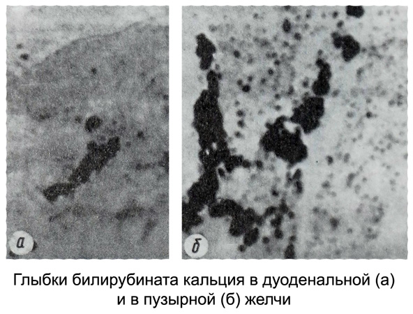
If you change the stability of the colloidal calcium bile bilirubinate can be found, along with cholesterol crystals and microlites.
Parasites and bacteria in bile
Vegetative forms of Giardia sometimes detected in all portions of bile. The fresh bile they are mobile, but on standing it become immobile. Giardia cysts are found in the stool. The value of giardiasis occur in the disputed cholecystitis. It is considered, that it supports the inflammatory process in the biliary tract and gall bladder.
Helminth eggs It can be detected in the bile in the liver helminthoses, gallbladder and duodenum (opistorhoze, fascioleze, klonorxoze, dikrocelioze, strongiloidoze, trihostrongilidozah).
To identify bacteria bile, collected into sterile tubes, sent for bacteriological examination (normal bile does not contain microorganisms). In these cases, it takes a special probe, in that in the region 0.2- 0,25 m from the mouth end of the glass tube is inserted. During the capture of a given batch of bile rubber tube is removed from the glass tube, end it is fired, and bile was collected in a sterile tube.
