Microscopic examination of the contents of the stomach
To prepare the formulations of the stomach contents in a thin layer of poured petri dishes, view on a black and white background, spatula and collected on a dissecting needle slides mucous, spotting, plotnovata pieces. It is necessary to study each of the three portions to prepare a preparation: native, Lugol solution dyed to detect starch and colored with Sudan III for detecting fat droplets. Material, supported on slides, covered with cover glasses.
Microscopic examination of the stomach contents to be, received an empty stomach and after the application of enteral and parenteral methods of stimulating the secretion of gastric.
At the same time found the elements of the gastric mucosa (slime, blood, epithelial, cloth scraps), food items during stagnation (corn starch, yeasts, lipids, muscle fiber) and microorganisms (sartsiny, Sticks lactic fermentation).
The mucus in gastric contents
The contents of the stomach is usually detected a small amount of mucus, stringy Klochkov, sometimes mixed with elements of food at a standstill or stomach epithelial cells and leukocytes. In atrophic gastritis mucus reduced, sometimes it can be completely absent. With increased gastric secretion microscopic slime almost impossible to detect due to its digestion. A significant amount of mucus in the gastric contents observed in hypertrophic gastritis and ahilii.
Also mucus, detachable gastric mucosa, misses in stomach contents from the mouth mucus and respiratory, in which a lot of air, so that it floats on the surface. Diagnostic value of this mucus has no.
Under the microscope, in the acidic gastric contents fibrous goo, and low acidity and ahilii - homogeneous. Detection planar in the mucus epithelial indicates its origin from the mouth. Identification of alveolar epithelial cells, often with a carbon pigment, indicates, it phlegm.
White blood cells in gastric contents
Leukocytes in the normal gastric contents are in the mucus, which partially protects them from the destructive action of hydrochloric acid and pepsin. First of all to digest their cytoplasm, however in the acidic gastric contents are presented as leukocytes naked nuclei, consisting of two or three segments (core Yavorsky). In the gastric juice with low acidity, especially when ahilii, the structure of white blood cells preserved.
The increase in the number of white blood cells with organic lesions of the stomach substantially has no diagnostic value, as it occurs in various functional disorders in the body. Excessive increase in the number of white blood cells observed in purulent inflammation of the stomach (gastric abscess).
Blood in gastric contents
The presence of blood in the stomach contents indicated by the appearance of brown pigment - hemosiderin (the color of coffee grounds). Microscopic examination of these cases, the red blood cells are not found, but sometimes found their shell.
At low acidity ahilii admixture of blood or stomach contents gives a reddish (bloody) tint, and microscopy revealed unmodified erythrocytes in mucus or without.
A small admixture of blood can be random, caused by mechanical trauma in probing
The presence of blood in the mucus scraps in the form of diffuse or brown pigment epithelial cells together with red blood cells of the gastric mucosa can be the result of hypertrophy of the gastric mucosa or deep lesions, accompanied by hemorrhage.
Epithelial cells in the gastric contents
Epithelial cells of the gastric mucosa are detected separately and clusters together with leukocytes in shreds of mucus. In the acidic environment of the stomach epithelial cells have the form of oval (round) cores, near. This is different from the nuclei of epithelial cells of flat polygonal epithelium of the mouth and esophagus, which, due to the large size of the cells is significantly distant from each other. The gastric contents with low acidity of gastric mucosal epithelial cells retain the cylindrical shape.
In hypertrophic gastritis gastric epithelial cells found in clusters, clusters, layers and casts of gastric glands, sometimes in large quantities, in krovjanisto-stained scraps plotnovatnyh. Sometimes they are subject to the mucosa and fatty degeneration, and metaplasia, whereby the rounded and flattened. A large number of epithelial cells of the gastric mucosa revealed by hypertrophic gastritis, and especially in the inflammatory process in the pyloric part of the stomach.
When polyps stomach They found the same type of layers of columnar epithelium with signs of proliferation: di-, tricyclic cells with enlarged nuclei, some of them are nucleoli.
In gastric cancer ability to detect plotnovata scraps of atypical epithelial cells and mucus, It is sometimes arranged in groups and formations zhelezistopodobnyh, frequent vacuolar or steatosis.
If chlamydia You can find Berezovsky-Sternberg cells, tuberculosis - Giant cells Pirogov-Langhans, Lumpy - Druze actinomycetes.
Elements of food in the gastric contents
Microscopic examination of stomach contents can be identified starch grains, yeasts, lipids and myofibers.
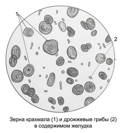
Corn starch - Part of bread, Potato, pulses and some other products.
Splitting of carbohydrates is influenced by the enzyme amylase (ptyalin), activated in an alkaline medium. This starch is first split into amidulin or amilodekstrin - soluble starch, which in the presence of iodine stained violet. The next step in cleavage is the formation of erythrodextrins., which are stained with Lugol's solution in a reddish color. Finally, achrodextrins and maltose appear, not stained with iodine.
The breakdown of starch stops in the stomach, t. it is. in an acidic environment. The higher the acidity, the earlier amylase is inactivated. With achlorhydria, the breakdown of starch continues to achrodextrins and maltose..
Thus, with increased acidity of gastric contents, purple-colored grains of starch are detected (amidulin), with normal acidity - grains are predominantly reddish in color (erythrodextrin), with achylia, as a result of the appearance of achrodextrins and maltose, staining is not observed, most of the starch is completely broken down.
yeast mushrooms – oval, strongly refracting light formations of spherical shape, forming characteristic budding structures (eights). When added with Lugol's solution, they turn yellow.. Yeast fungi, together with starch grains, are detected in the gastric contents after a bread trial breakfast or are detected during congestion in the stomach.

Neutral oil has the form of drops of various sizes, which are stained orange-red with Sudan III. Occurs with congestion in the stomach.
Muscle fibers in the gastric contents of a patient with low acidity, they look like cylindrical formations of a yellowish or greenish color with a characteristic transverse striation. Their presence indicates congestion in the stomach..
Microorganisms in the stomach
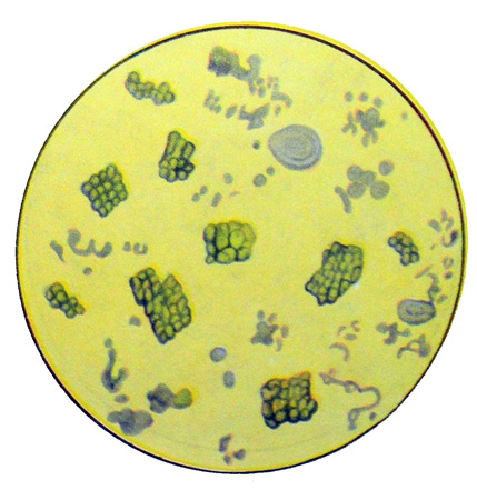
Microscopy of gastric contents sometimes reveals microorganisms, in particular sarcinas, Sticks lactic fermentation.
Sarcins – cocci, which are divided in three mutually perpendicular planes and look like tied bales.

They can be detected with delayed evacuation of food with the presence of free hydrochloric acid in the gastric contents..
Lactic fermentation sticks (Boasa-Oplera) - long, rough, slightly curved, often at an angle. Occurs with delayed evacuation of food in the stomach in the absence of free hydrochloric acid.
