Microscopic examination of stool: what is this analysis, how and why is it done
Microscopic examination of the stool can reveal detritus, leftover food, elements intestinal mucosa, Crystals, organisms.
Detritus It represents the remains of food items, Microorganisms, disintegrated rejected by gut epithelium, leukocytes, erythrocytes, etc.. It looks fine grained amorphous formations mainly form. As the detritus of the bulk of feces, the greatest number found in his stool decorated and smallest - in liquid. The thinner cal, the smaller detritus. By quantity of detritus can be judged on digestion. When making these microscopic examination of the nature of detritus not say.
Slime. On gross examination of stool mucus can not find, since it covers the surface of normal feces thin, barely noticeable layer. Microscopic mucus is detected as a structureless substance with a single cylindrical epithelium cells.

Increasing the amount of mucus in the stool in adults indicates a pathological condition. Newborn small flakes of mucus found in physiological conditions.
Epithelial. The stool can detect cells flat and cylindrical epithelium.
Squamous cells of the anal canal are located in isolation or layers. Finding them has no practical significance.
Cylindrical epithelial fall into the feces of all the departments of intestines. They may be unchanged or undergo degenerative changes. In the latter case, epithelial cells shriveled, reduced, waxy, sometimes denuclearized, may take the form of matt grain.

There are epithelial cells in the mucus of the large intestine. Normally in the stool contains a small number of cells of columnar epithelium. When catarrh intestinal mucosa epithelial cells can be detected in a large number of individual cells and entire layers. The ribbon-like films in the mucous colic (pereponchatom cars) Cylindrical epithelial also possible to identify a large number of.
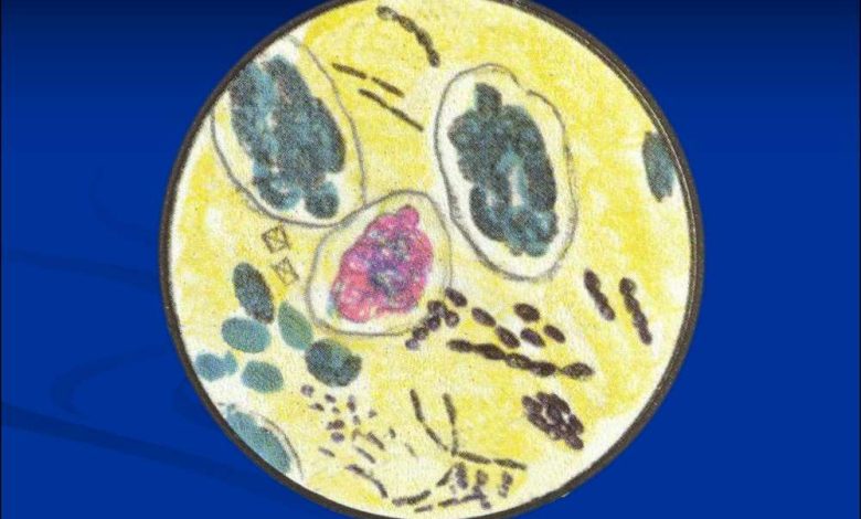
White blood cells, predominantly neutrophilic granulocytes, They are either in the mucus, or is it. When catarrh intestinal mucosa a small number of white blood cells, ulcerative process, it increases sharply, especially if it is localized in the distal intestine.
Eosinophilic granulocytes are observed in spastic colitis, amoebic dysentery, Some helminthoses. When added to the mucus 5 % aqueous solution of eosin stained grain of bright orange. Often along with eosinophilic granulocytes found Charcot-Leyden crystals.
Macrophages found in stained, various sizes, most major, with round nuclei, they are located in the cytoplasm of inclusion: erythrocytes, neutrophilic granulocytes (whole or fragments). In dysentery macrophages found in small amounts, when amebiasis - isolated.
Red blood cells identified or unchanged, or in the form of shadows, which are difficult to detect. They can be released in the feces and in the form of amorphous decay, painted in brownish color. The presence of red blood cells indicates, usually, the presence of ulcer process. Unmodified red blood cells are usually found in the stool bleeding from the lower gastrointestinal channel (hemorrhoids, rectal cancer, and others.) and heavy bleeding from upper alimentary canal. Sometimes the red blood cells are detected in the feces with mucus.
Vegetable fibers present in feces is constant and often in a large amount, due to constant use plant food.

Digestibility of vegetable fibers chemical composition refers to polysaccharides. It is composed of cells, with tender, thin, easily destroyed by shell. Digestive enzymes can easily penetrate the cell membrane of digestible fiber, even if it is not damaged, and cleave their contents.
Cells of fiber interconnected layer of pectin, which initially dissolves in the acidic contents of the stomach, and then weakly alkaline contents duodenum. When ahilii digestible fiber cells do not separate and are found in feces in groups (Potato cells, carrots, etc. I.). Decorated feces digestibility of fiber is absent.
The indigestible vegetable fiber nakhoditsya lignin, giving it hardness and rigidity. Cells indigestible fiber membranes have a thick double-circuit. The human digestive tract does not produce enzymes, ability to cleave membrane of plant cells. Fiber helps splitting some microorganisms of the large intestine (clostridia, Bcellulosae disssolvens and others.). The longer the stool is in the guts, the less fiber it is. The structure of the indigestible plant cellulose is very diverse, most characteristic of her presence of residues of leguminous plants in the form of narrow, long, parallel palisade cells, refracting light; vascular plants, spiraley, hair and needles, epidermal cereals etc..
Corn starch It is found in the feces of extracellular and cell potatoes, bean, etc.. d. They are easily identified by the addition of iodine.

Corn starch, located extracellularly, lose their bedding and have the form of irregular rubble. Depending on the stage of digestion of the starch grains adding Lugol stained differently: amilodekstrin gets purple color, eritrodekstrin - red-brown; coloring does not change arhodekstrina. Normally starch grains in the stool are missing. Incomplete breakdown of starch occurs in diseases of the small intestine and the associated rapid evacuation of food.
Muscle fibers. Remains of protein foods in the form of muscle fibers can sometimes be identified even at the macroscopic study of feces. Microscopic examination of the remains of the muscle fibers are found in any drug, even if the patient is taking the food with a small amount of meat.
Overcooked muscle fibers have the form of ovoid neischerchennyh debris of various sizes. Insufficient digested fiber longitudinally striated, of the acute angles. In unaltered muscle fibers maintained transverse striations, all sharp angles.

If insufficient flow of bile into the duodenum muscle fibers pale-colored. Under the influence of hydrochloric acid gastric juice muscle fibers are released from foodborne intermuscular connective layers and sarcolemma. This violates the structure of muscle fibers, of transverse and longitudinal striations. In this state, most of the muscle fibers enters the duodenum. The final digestion of the muscle fibers is mainly influenced pancreatic juice. The appearance in the feces of a large number of groups of muscle fibers with preserved transverse and longitudinal striations indicates insufficiency digest food in the stomach.
A large number of muscle fibers (kreatoreya) It may be due to:
- axilii (the presence of drugs of striated, or striated, muscle fibers);
- insufficient pancreatic secretion (presence in the formulation and insufficiently sufficiently digested, separately arranged muscle fibers);
- abnormally rapid evacuation of food (the presence of undigested fiber);
- alimentary overload, which should not be after the test diet. What matters it is also a method of cooking meat and condition of the masticatory apparatus.
Connective tissue. Calais, highly diluted with water, connective tissue particles have the form of fragments and strands of grayish color of irregular shape with shaggy torn edges. On microscopic examination, they are characterized by a delicate filamentary structure, but from mucus are more sharp outlines, more dense texture and opacity. After adding acetic acid connective tissue structure disappears, and mucus appear stratification and striation. When eating poorly cooked meat and roasted presence in the feces of the connective tissue - a physiological phenomenon.
Detection of connective tissue after a trial diet (especially diet Schmidt) evidence of failure of digestion of food in the stomach.
Fat. Normally cal always contains a small amount of fatty acids and their salts. Neutral fat is absent.
The native preparation neutral fat takes the form of round or oval colorless or slightly yellowish drops. When pressed on the cover glass drops alter the outlines. If a lot of fat, they merge. The preparation, stained with methyl- new blue, colorless drops of neutral fat, and treated with Sudan III - bright red.

Fatty acids found in the feces in the form of long, pointed needles (Crystals), sometimes stacked in bundles, as well as in the form of drops and lumps, sometimes with spikes.

If you find a needle and lumps in his native preparation is heated, not boiling, and examined under a microscope. Fatty acids when heated to form droplets, that, cooling down, again turn into lumps. Warming can be repeated many times. Drops of fatty acids stained with methylene blue blue.
Soap (salts of fatty acids) It occurs as clumps and crystals, Crystals similar to fatty acids, but shorter, often located beams.

If the drug is heated needles and lumps do not form droplets, the drug must be heated with acetic acid (20-30 %) to boiling. Education drop indicates the presence of soaps: acetic acid cleaves and releases the soap fatty acids, which melt, forming drops.
If the digestion of fat the most important role played by lipase pancreatic juice and bile. Violation of pancreatic secretion causes, that the fat is not digested and excreted in large numbers in the faeces. If no bile enters the duodenum, the fatty acid, formed from neutral fat by lipase, not absorbed and are present in the stool in large numbers. Feces with significant fat (celiac disease) It has a unique pearlescent, grayish color and consistency ointment. It can be detected and pieces of undigested fat tissue. This is observed in case of violation of digestion in the stomach, where normally fat is released from the connective tissue.
Crystals. Tripelfosfaty as crystals occur most frequently in the liquid excrements and mucus. The reaction with the alkaline feces. Diagnostic value of detection is only freshly feces. Usually, the appearance of these crystals is associated with increased putrefaction in the feces and urine admixture to it.
Oxalates found in the feces when receiving a large number of plant food. Normally, hydrochloric acid converts calcium oxalate in calcium chloride, so the presence of oxalate in the stool may indicate a reduced gastric acidity.
Cholesterol crystals fecal recognized with difficulty and do not have a diagnostic value.
Charcot-Leyden crystals observed in the feces when ingested it eosinophilic granulocytes. When these crystals amebiasis sometimes reach large sizes.
The crystals of bilirubin can obnaruživatʹsâ in profuse proud, When bilirubin is not time to recover stercobilin due to rapid evacuation of food through intestines. They represent a small pointed at both ends needle-like crystals yellowish brown, beams arranged in a.
Crystals gematoidina appear in the stool after intestinal bleeding in long needles and rhombic plates. Their color varies from golden yellow to brownish-orange.
Microflora. The human colon is a large number of microorganisms. They constitute 40-50 % stool weight and are part of detritus. The practical significance is the discovery in feces iodophilic flora and Mycobacterium tuberculosis.
K jodofilʹnoj flora are micro-organisms (cocci and rods of various lengths and thicknesses), having the property of staining solution Lugol black due to the presence of these granulosa. Iodophilic flora growing on media, containing carbohydrates, she assimilates.
Under physiological conditions iodophilic Flora is located in the lower part of the ileum and blind guts. Normally, the content of it in the stool is very small, and constipation is absent. An increase in the fecal flora iodophilic combined with acidic, accelerated release of chyme from the intestines and the emergence of fermentation processes. When expressed fermentation processes found in the feces long, slightly curved sticks, which are located in groups and chains - leptotriks and thick spindle-shaped bacilli, sometimes with swelling at one end (a drumstick) - clostridia, forming groups and chain, sometimes lying intracellularly. Clostridium stained with iodine or entirely, or only in the middle part.
If the fermentation is expressed mild and combined with the process of decay, can be found in the feces of small cocci and rods. Yeast fungi are stained with a solution of Lugol yellowish. Finding them in large quantities in fresh feces indicates candidiasis.
Mycobacterium tuberculosis are found in the feces of tuberculosis of guts. Preparations for the study of special prescription made from the mucous, muco-purulent and bloody lumps, in the absence of mucus, blood, pus - Stir of water stool, fixed and stained by Ziehl-Nelsenu.