Macroscopic examination of stool: analysis, research to diagnose disease
Macroscopic examination of stool includes inspection, determination of its amount (if necessary), colors, consistency, shape, odor, reaction and visible impurities (food particles, mucus, pus, parasites, etc.).
Number of feces per day It depends on the amount of ingested food, her character, digestibility, peristalsis of the intestines, and the amount of water. Normally, on average, 100-200 g of feces (which is 30-35 g dry substance), when plant foods - up 400 g and more.
In case of violation of digestion (Akhil stomach, enteritis, pancreatic lesion et al.) We observed a significant increase in stool. Especially increases the mass of faeces (to 1 kg per day and higher) while pancreatic disease and sprue.
Painting feces It depends on the presence of stercobilin and the nature of the food. Normally, the color yellowish feces- or brown-brown, with exclusively milk diet - yellow or light yellow. Sometimes the color of feces inhomogeneous: along with dark brown found the lighter particles.
Eating cherries, Other products, containing natural dyes, It causes darker stool.
Plant food, rich in chlorophyll, feces stains green or greenish-brown color.
Change the color of feces, some drugs (Carbolite, iron supplements, bismuth, arsenic).
Blood in different amounts can also change the color. When bleeding from the stomach, duodenum and upper small intestine stool is black, tarry. When bleeding from the distal portions of the small intestine to the color of feces mixed with a reddish tint, and in the case of bleeding from the colon becomes red feces, bloody color. The color of stool is dependent not only on the site of bleeding, but also on the amount of blood streamed.
Color clay stool often has impurities at high fat. In the absence of bile there aholichny (unpainted) mud.
The consistency of the feces OK soft, in pathological conditions - dense (constipation), mazevidnaya (with significant impurity fat), pappy, semi-liquid or liquid. Density depends on the stool water content. Normally found in the feces 80 % water. In the presence of 75 % Cal Water tight, 85 % - Pappy, 90 % - Liquid.
Contents water Calais due to the length of stay of stool in the intestines. With persistent constipation stools it is particularly long in the intestines, so there is less water, and it is tight. In ystoschennыh, malnourished people feces dry and hard. With the accelerated evacuation of the water content of the food in the stool increased, it is less dense. Usually in such instances allocated unformed, kashitseobraznыy mud. With increasing fermentation processes semi-stool can have a foamy character. Often at the same time stand decorated and liquid excrements. In acute enteritis and cholera watery feces.
Shape of feces. In a healthy person usually allocated cal cylindrical shape. With increasing density, it takes the form of lumps of various sizes. Most lumps are seen on the surface of the constriction of the large intestine. With spastic conditions, when feces especially long stays in the intestines, it lumps are very small and resemble sheep feces. When muscle spasms of the rectum and colon tumors may have a ribbon-like stool form.
The smell of feces It depends largely on the presence of degradation products of protein foods: skatol, indola, less phenol, etc.. He sharply when meat food, than vegetable.
When pride latch kala It may be putrid, when decaying malignant colon tumors mud zlovonnыy (the smell of carrion).
When expressed during fermentation in the gut sour smell of faeces on the presence of volatile fatty acids (oil, acetic et al.).
In the event of prolonged constipation faint smell of feces, fasting and he did not.
Kal infants almost odorless or with a slightly sour smell. Odor of feces in infants has a diagnostic value.
The reaction of feces determined by the change in color of the indicator paper universal, pre-wetted with distilled water.
Normally, when using mixed meal fecal neutral reaction (pH 7,0) or slightly alkaline, and when the food, rich in carbohydrates,- Weak acid. When enhancing the processes of decay reaction feces usually rezkoschelochnaya, and with enhanced fermentation - acid.
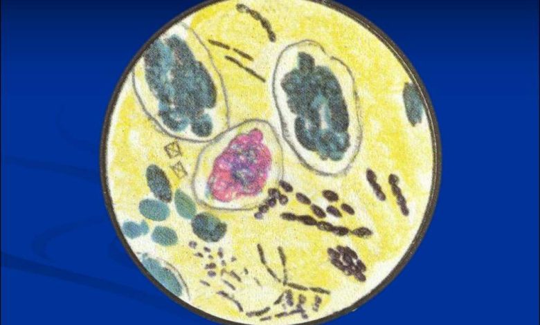
The remains of undigested food visible to the naked eye. In liquid feces can be detected immediately, in a dense and mushy - after dilution with water. To this stirred cal, part of it is ground in a porcelain mortar, or a petri dish with water or isotonic sodium chloride solution, whereupon the material is taken for microscopic examination. It is possible to identify the remains of food (slices of meat, scraps of connective tissue, the remnants of fat and fiber), slime, blood, pus, individuals and segments of helminth, concretions, scraps of fabric.
Slime Cal usually covers decorated with a thin film, so that its surface is slippery, a slightly shiny. The amount of mucus increases considerably in inflammatory processes in the gut. Some increase in mucus in the stools with constipation is a defensive reaction to the irritation of the intestinal mucosa dense lumpy masses. Mucus may be on the surface of the stool, or mixed with it.
The nature of the location of the mucus in the stool and its amount have great diagnostic value. The smaller lumps of mucus, and the closer they are mixed with the feces, the higher place of their allocation. The consistency is viscous mucus, soft or dense. It can be colored differently. The most common color is gray-whitish mucus, may appear pink (reddish) tint or coloration bile into the small intestine yellowish or yellowish-green color. Flakes mucus, painted in yellow color, indicate the failure of the small intestine.
Normally, the mucus of the small intestine has time to digest, so its presence in the stool indicates a rapid gut peristalsis. Sometimes a lot of mucus, and it stands out as a ribbon-like film, Recalling appearance tapeworms. Such films are in the mucous colic (pereponchatom cars). With spastic colitis mucus is in the form of lumps on the surface of the stool, or between lumps.
For the detection of mucus in his unformed stool was diluted with water in a petri dish or isotonic sodium chloride solution and browsing on white and black background: on white lumps mucus transparent than the surrounding feces, and in black - darker than the surrounding fluid. With a small content of mucus can be found only under a microscope. To identify its use dyes. Reagent Gehta (mixture 0,2 % and brilliant green 1 % neutral red in equal volumes) mucus gives a reddish tint, and feces stains green. Triatsid Ehrlich stain the mucus in the blue-green color.
Fecal blood there may be bleeding from different parts of the alimentary canal. Blood clots or diffusely stained with blood feces observed with haemorrhoids, yazvennom cars, chronic constipation, sigmoid colon polyps, rectal cancer, fissures, etc.. The blood may be mixed with mucus.
Pus excreted in the feces with dysentery, tuberculosis, ulceration of the distal small intestine, decay cancer, paraintestinalnogo breakthrough abscess, etc..
Worms. When helminths in stool can be detected by special round worms and tape segments.
Stones. Perhaps revealing bile, pancreatic and fecal stones. Gallstones can be cholesteric, Bilirubin, calcareous, mixed. Pancreatic - small size (pea), porystыe, composed of calcium carbonate or calcium phosphate. Fecal stones, or coprolites, It consists of tightly compacted feces (mostly from plant fiber, impregnated with calcium salts) and can reach the size of a walnut.
Pieces of tissue may be in the feces with dysentery or decay of cancer.
Methods of macroscopic fecal
Macroscopically visible particles of feces (other than edible) select and prepare them for microscopic examination preparation.
Besides, Cala, diluted with water or isotonic sodium chloride solution, preparing four more preparation for microscopic examination, Why four slide glasses cause a drop of fecal emulsion and added to a solution of Lugol, to another - methylene blue, third - acetic acid (20- 30 %), and the fourth drug left the native. Content on slides stirred, covered with a cover glass and examined under a microscope.
For the detection of helminth eggs prepared drug stool with glycerol, which is added to detect Clostridium (iodophilic flora), yeasts, lamblia cysts and starch grains.
Methylene blue and acetic acid necessary for the differentiation of fat and their cleavage products. In the native preparation, the extent of digestion of food components (proteins, fat, carbohydrates).