White blood cells – Degenerative changes of leukocytes
Characterized by the deposition in the cells of various endo- and exogenous substances, as a result they lose their ability to function normally and division. Pathological material, shall be deposited in the cell and causing degenerative changes in white blood cells, It may be different in nature:
- lipids (zhirovaya degeneration);
- pigments (pigmentnaya degeneration) etc..
Of great importance in clinical practice is research as a morphological structure of leukocytes, and their functional status, the presence of degenerative changes.
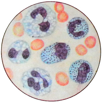
Degenerative changes of white blood cells are neutrophils grain toksogennaya (TMT), vacuolation, the appearance of cells Knyazkova-Case, zeren Amato, etc..
Toksogennaya granularity of neutrophils
Formation of specific grain begins on neutrophils Golgi. Sizes range from pellets 0,2 to 0,5 m, their rounded shape or slightly oblong.
Rounded, primary, grain contains immature cells neutrophil series - promyelocytes; oblong, secondary, specific - mature neutrophilic granulocytes - and stab-core segments.
The myelocytes and metamyelocytes detected as primary, and secondary granules.
Education toksogennoy grain It occurs within the cell as a result of physicochemical changes in the protein structure under the influence of the cytoplasm products intoxication. Its appearance is explained by the release into the peripheral blood neutrophilic granulocytes immature bone marrow, containing primary granules, rich in proteins, has antibacterial properties, and glycosaminoglycans and lysine.
Toksogennaya granularity of neutrophils often appears before the nuclear shift. Her rise in the septic diseases, lobar pneumonia and a number of inflammatory diseases indicates the progression of the pathological process and the possibility of an adverse outcome. In a lot of grit toksogennaya neutrophils appear in the decay of the tumor tissue under the influence of radiation therapy. Most expressed toksogennaya grain with lobar pneumonia during the resorption of inflammatory infiltrate, scarlet fever, septikopiemii, peritonitis, cellulitis and other purulent processes. Particularly important is the toksogennaya grain in the diagnosis of acute abdomen (eg, gangrenous appendicitis, accompanied with slightly increased body temperature and often in the absence of leukocytosis). In describing toksogennoy grain (blood smear stained blue by E karbolfuksinmetilenovym. ABOUT. Freyfeld) indicate the percentage of cells containing it and the size of grain (fine, medium size, large, flocculent).
Taurus, Dele Knyazkova
Taurus, Dele Knyazkova found in the cytoplasm of neutrophils in the form of rather large pale blue lumps of various shapes. There are inflammatory and infectious diseases, occurring even mild, when toksogennaya grainy still poorly developed or absent.
There are several points of view on the origin of cells Knyazkova-Case. According to one, they have nuclear origin, the other - are the remnants of basophilic spongioplazmy young cells, third -th same origin, that grain toksogennaya.
Zerna Amato
Grains Amato detected in the cytoplasm of neutrophils in the form of small rounded, oval or similar entities comma, painted in light blue color and containing red or red-purple corn. They appear in scarlet fever and other infectious diseases. By origin they are considered close to the calves Knyazkova-Case.
Vacuolation tsitoplazmы
An important feature of the degenerative changes in white blood cells is also vacuolation cytoplasm. It is found less often, toksogennaya granularity than neutrophilic granulocytes, but it is no less important diagnostic value, pointing to the severity of illness or intoxication. The most typical forms of vacuolation for severe sepsis, abscesses and acute liver disease.
In acute sepsis, caused by anaerobic infection, with severe leukocytosis (above 50 T in 1 l) vacuolization can be observed almost all leukocytes - "full of holes", "Riddled", leukocytes.
When counting leukogram or simply indicate the presence of vacuolization, or the number of cells with vacuolated cytoplasm as a percentage is expressed on 100 neutrophilic granulocytes, more properly.
The signs of degeneration are also hypersegmentation cores, xromatinoliz, karioliz, fragments, pyknosis, karioreksis, cytolysis etc..
The anomaly of leukocytes Pelgera
It was first described in the Dutch hematologist Pelgerom 1830 g. At the present time it is often. Inheritance of this anomaly is carried out by dominant pattern of one of the parents (heterozygotes) or, that is rare, from both (homozygotes).
In the blood, suffering from this anomaly detected a huge number of leukocytes krugloyadernyh. In theory, the probability of the homozygous variant pelgerovskoy anomaly is 1:1000000, in fact, it is even rarer.
We describe the case of a total of four significant homozygotes. When heterozygous inheritance anomaly is transmitted from generation to generation and is determined at the 50 % family. Thus, availability of individual family members does not contradict the normal leukogram family hereditary anomaly.
A feature of leukocyte anomalies Pelgera is the form of the nucleus.
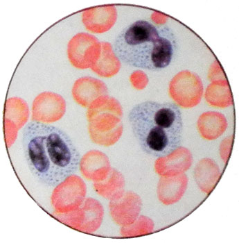
Most of neutrophilic granulocytes have odnodolevoe unsegmented ellipse, bean-shaped or kidney-shaped nucleus, shorter, than the usual core of neutrophilic granulocytes. In other cells, the nucleus with the emerging middle constriction shaped like a weight or gymnastic groundnuts (peanuts). There are also cells with nuclei, transition to dvusegmentarnym (having the form of eyeglasses); three core segments are almost never found.
How two-, and different forms of trisegmentoyadernye short jumpers and lumpy structure of the nucleus. Neutrophil granulocytes with a large number of segments at Pelgera anomalies do not occur. Along with non-segmented, stab and segmented neutrophilic granulocytes observed round-core, are recognized fully mature cells. The peculiarity of their development lies in the complete absence of nuclear polymorphism, t. it is. core structure of chromatin old, but in form - young.
In order to avoid erroneous interpretations of the analysis in the presence of these forms of neutrophilic granulocytes physician assistant is required to give an opinion on, that this picture is characteristic of the blood leukocyte anomalies Pelgera.
With such changes in the shape of the nucleus in eosinophilic and basophilic granulocytes, these cells are also counted differentially (krugloyadernye, non-segmented, stab, dvusegmentoyadernye and trisegmentoyadernye).
As white blood cells in the physiological properties of the anomaly Pelgera are no different from the usual. The women - carriers of this anomaly is not detected sex chromatin. Women - partial media anomalies can be observed neutrophilic granulocytes with sex chromatin (the latter in the form of Taurus Barra It is found in the nuclei of cells of the oral mucosa, which typically investigates sex chromatin). This phenomenon is due to a delay segmentation nuclei pelgerovskih neutrophilic granulocytes, whereby the sex chromatin is as if immured in the mass of the nucleus.
The bone marrow is dominated by neutrophilic granulocytes krugloyadernye (to 65 %). Among them are found mature cells with round, oval or ellipsoidal nucleus. In the same way is the development of characteristic abnormalities Pelgera krugloyadernyh eosinophilic granulocytes. Erythrokaryocytes are no more than 15-20 %, and found almost exclusively normoblasts with pyknotic nucleus at different degrees gemoglobinizatsii.
Along with the anomaly of leukocytes Pelgera, nosyashtey family-nasledstvennыy character, In recent years, the description of acquired forms giposegmentatsii nuclear neutrophilic granulocytes - pelgeroidov.
Psevdopelgerovskie leukocytes, unlike true, Blood found impermanent. Their appearance is due to the underlying disease.
It should also be aware of the possible presence of abnormalities in carriers of both pelgerovskoy anomalies of leukocytes Shtodmeystera. Unlike typical pelgerovskih krugloyadernyh neutrophilic granulocytes with gruboglybchatoy fragmented structure of the nuclei, with clear contours, kernel when anomalies are characterized by less pronounced Shtodmeystera chromatin condensation, presence buhtoobraznoy notch and unique fringed, consisting of delicate threads of chromatin, like projecting from the main mass of the nucleus into the cytoplasm. This anomaly also runs in families and can be found not only in the form pelgerovskom, but also independently.


