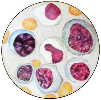
Leukemoid reaction
By leukemoid reactions include changes in the peripheral blood and blood-forming organs, similar to leukemia, but in contrast with reactive. Leukemoid tests never go in leukemia.
There are leukemoid reaction myeloid and lymphatic types.
Besides, in 1979 allocated psevdoblastnye leukemoid reaction, are observed in patients at the outlet of agranulocytosis and characterized by the appearance in the peripheral blood and bone marrow blast cells with a homogeneous core, containing one or two nucleoli, and a narrow rim of cytoplasm grain-free.
Relative lymphocytosis and lack of transitional cell granular in peripheral blood can cause suspicion of acute leukemia. However, short-term stay (about a day) these cells in peripheral blood and the absence of a detailed study of their characteristic structural features of blast cells to rule out this diagnosis.
Reactions myeloid type
These reactions include promyelocytic, neutrophilic shift to the left, the reaction of two or three of germs myelopoiesis, monocytic and reactive cytopenia - Watering- thrombocytopenia and.
Promyelocytic reaction
Promyelocytic reactions can occur when agranulocytosis, after the appearance of blast forms.
The large number of promyelocytes with abundant grain resembles acute promyelocytic leukemia. However, the lack of hemorrhagic syndrome, characteristic of acute promyelocytic leukemia, thrombocytopenia, polymorphism of nuclei and grain promyelocytic, Negative reaction to the sulfated acidic glycosaminoglycans to rule out this diagnosis.
Promyelocytic leukemoid reaction in the bone marrow may be due to poisoning, atopic dermatitis as a result of medication, and other factors influence. In all these cases, there are no specific clinical and morphological features as the promyelocytic leukemia, Tac and agranulocytosis.
Promyelocytic leukemoid reactions may occur in hereditary neutropenia. Monocytosis and eosinophilia in the bone marrow, and sometimes in the blood, specific to hereditary neutropenia, never occur in acute promyelocytic leukemia.
Neutrophil reaction shift to the left
Neutrophil reaction shift to the left until promyelocytic observed in inflammatory and suppurative processes, sepsis and chronic myeloid leukemia. Start of chronic myeloid leukemia usually asymptomatic, whereas inflammatory, suppurative processes, and there is always a sepsis clinical symptomatology.
Neutrophilia with a shift to the left occurs in acute hemorrhage, However, in cases of accession poisoning, when expressed mild leukocytosis possible significant stab shift 30-40 % without metamyelocytes and myelocytes. In the bone marrow at the same time significantly increasing the number of promyelocytic, the ratio of white blood cells and red blood cells dramatically increases due to the first. In contrast, chronic myeloid leukemia, in reactions of this type leukemoid no increase in platelet count.
Reaktivnaя эozinofiliя
Reactive eosinophilia as a manifestation of sensitization of the organism It can be observed in helminthiasis, systemic connective tissue diseases, Cancer, chlamydia and other diseases. In chronic myelogenous leukemia eosinophilic granulocytes, along with other granule cells, is a substrate of the tumor.
The reaction of two or three of germs myelopoiesis
The reaction of two or three myelopoiesis germs can occur in cancer metastases to bone marrow.
There are two types of reactions leukemoid:
- the first is characterized by neutrophilic leukocytosis (often with a pronounced shift to the left, sometimes with a slight increase in the number of basophil granulocytes), thrombocytosis and erythrocytosis less;
- for the second characteristic neutrocytosis (shift to myelocytes) and the appearance in the peripheral blood of different maturity erythrokaryocytes.
When multiple metastases in the bone marrow develop anemia and thrombocytopenia. The number of white blood cells varies from leukopenia to a small leukocytosis. The blood smear - neutrophilia with a shift to the left to myelocytes and promyelocytes, find erythrokaryocytes (This type of reaction resembles leukemoid erythromyelosis).
The same pattern can occur in acute immune hemolysis, which is characterized by leukocytosis with a shift of neutrophils to promyelocytes and myelocytes, there may be individual normoblasts. In peripheral blood there are reticular cells, amount in the bone marrow they can reach 15 %.
Reticulocytosis in peripheral blood, jaundice, urobilin significant amount in urine, hemoglobinuria and gemosiderinuriya, anemia, irritation of the germ in red bone marrow in the absence of increase in the number of blast cells to rule out acute erythromyelosis, at which hemolysis may also be observed, but he is not a leading symptom of the disease, as in hemolytic anemia.
Monocytic reaction
Monocytic reaction reminiscent of chronic monocytic leukemia, develops in people over the age of 50 years, and occurs without clinical manifestations over the years. Reactive monocytosis appear in the background of a disease. Most often they occur in tuberculosis, rheumatism, toxoplasmosis, hereditary neutropenia, paraproteinemic hemoblastoses, chlamydia and other diseases.
When difficulties in the diagnosis of bone marrow biopsy shows. When monocytic leukemia in trepanate dominated tumor cells (large cells with large light nuclei, nucleoli, and often pale blue cytoplasm).
Reactive cytopenia
These include acute lejko- thrombocytopenia and, developing as a result of the rapid consumption of a large number of cells (cytopenia consumption).
Reactive cytopenia, especially leukopenia, are rare. They arise usually after cytostatic therapy as a result of exhaustion or depression in these patients bone marrow granulocyte reserve (eg, in cancer patients).
Thrombocytopenia can occur after consumption of infectious shock, thrombosis, DIC with massive consumption of platelets and decrease in their number in peripheral blood. Perhaps the appearance of thrombocytopenia consumption after hemorrhage. Sometimes thrombocytopenia can be accompanied by leukocytosis with neutrophilic shift to the left and anemia.
To differentiate cytopenias consumption and aleukemic stage of acute leukemia (if necessary) investigated of bone marrow (if cytopenia consumption it lacks cell acute leukemia).
The reactions of the lymphatic type
For reactions of this type include oligosymptomatic infection lymphocytosis, reminiscent of the pattern of blood chronic lymphocytic leukemia, and infectious mononucleosis, which often have to be differentiated from acute leukemia.
Malosimptomno infectious lymphocytosis
Malosimptomno infectious lymphocytosis is characterized by high leukocytosis expressed by peripheral blood lymphocytosis mild clinical symptoms or no. The most commonly occurs in children between the ages of 2 to 7 years, although the occurrence of the disease in two months of age, and in people older 17 years (to 25).
It is believed, it is a viral disease, wherein the pathogen enters the body through the mucous membranes of the nasal part of the pharynx or the digestive tract. The disease is characterized by considerable contagiousness.
Clinical symptoms very sparse and appears briefly (12-24). The onset sudden or gradual, It can manifest abdominal pain and other symptoms of enterocolitis. Occasionally develop meningeal symptoms. Most of the patients suffer from a lack of appetite, rapid fatigue. Often, the body temperature rises, It appears nasopharyngitis.
In some cases, the period of the rise of symptoms goes unnoticed. Duration of disease, according to different authors, It ranges from a few weeks to three months or more.
Peripheral lymph nodes are not normally increased, but occasionally, a slight increase in cervical lymph nodes, sometimes the tonsils. Described isolated cases with a slight increase in spleen.
An important diagnostic feature of the disease is hyperleukocytosis with lymphocytosis. The number of white blood cells ranges from 40-60 to 100-150 F in 1 l. The hemogram lymphocytes are 80-90 %. This cell medium and small sizes, which are characterized by dense structure of the nucleus and light blue narrow cytoplasm. Dual-core found lymphocytes and reticular cells. The number of erythrocytes and thrombocytes remains unchanged.
The dramatic increase in peripheral blood lymphocytes with no or insignificant hyperplasia of the lymph nodes, apparently, It may explain the increased elimination of lymphatic cells from lymphoid organs into the blood, which is confirmed by histological study sections. Established, that at malosimptomno infectious lymphocytosis lymph nodes lymph follicles are small, some of them hyalinized. This is due to the action of lymphotropic virus.
Qualitative and quantitative changes in the bone marrow are not found. The increase in the number of lymphocytes in the myelogram is a consequence of dilution of bone marrow punctate peripheral blood.
Blood picture in infectious lymphocytosis reminiscent of chronic lymphocytic leukemia. However, infectious lymphocytosis usually develops in children and less often in adolescence, and chronic lymphocytic leukemia - the elderly.
Kissing disease
Infectious mononucleosis - a disease, caused by a virus, has a tropism to the system of mononuclear phagocytes, and especially to the lymphatic tissue.
This disease was first described in 1885 g. Russian pediatrician H. F. Filatov. Among the acute lymphadenitis, he singled out an independent nosological form under the title "idiopathic lymphadenitis". IN 1889 g. Pfeiffer described it as a disease called "glandular fever". There are many synonyms of names of the disease, However, the conventional current and reflects the essence of the process is the morphological "kissing disease".
Develop infectious mononucleosis in all age groups, but most often - in adolescents, boys and young people. Most suffer from males. The incidence increases in autumn and spring.
Symptoms of infectious mononucleosis
The disease can begin with the appearance of the main clinical symptoms:
- Fever;
- Sore Throat;
- Lymphadenopathy;
- Splenomegaly - or only one of them,.
Fervescence usually preceded by a sore throat and lymphadenopathy. Sore throat observed in 90 % patients with infectious mononucleosis and can be bluetongue, necrotizing and psevdodifteriticheskoy. Lymphadenopathy in infectious mononucleosis - an earlier and permanent symptom, than angina, and only in rare cases, it appears after the sore throat.
Size of lymph nodes sizes ranging from bean to walnut or more. Sometimes they are barely be felt or is not increased. The most characteristic swelling of the lymph nodes in the posterior edge of pectoral-clavicular-mastoid muscle; increase as anterior cervical submandibular, axillary, inguinal lymph nodes and other
You can zoom in mediastinal lymph nodes and abdomen. The largest size they reach to the 4-6 th day of the disease, 10- to 15-th usually decrease in size, however, slight swelling of the lymph nodes can be seen for weeks or months. The consistency of the lymph nodes elastic, they are not fused with each other and with the skin, slightly painful on withdrawal of the fingers during palpation.
Splenomegaly, usually, insignificant. There are cases splenomegalic form of infectious mononucleosis. Reducing the size of the spleen to normal very slowly. Increased liver 10 30 mm is observed in most patients with infectious mononucleosis. In some cases there is jaundice.
The above clinical picture is observed in approximately 50 % cases. In other patients the process is characterized by a wide variety of clinical symptoms.
Diagnosis of infectious mononucleosis
The diagnosis of infectious mononucleosis placed on the basis of morphological examination of blood. The main signs of hematologic diseases are moderate leukocytosis and the predominance of mononuclear cells and atypical forms in peripheral blood.
Leukocytosis with infectious mononucleosis varies 10 - 20 T in 1 l, However, a more significant increase, and cases, proceeding with leukopenia. In young children, leukocytosis above, than in older children and adolescents. The most significant leukocytosis observed between the 5th and 12th days of illness. Duration leukocytosis can be from two weeks to several months.
The number of lymphocytes in the hemogram or in the normal range has been increased to 50-70 % by medium and large, shirokotsitoplazmennyh, lymphocytes. There a small monocytosis, but possibly normal amount of monocytes.
Of particular interest are atypical mononuclear cells, which in some cases may be numerous and diverse.

The size of these cells range from medium to large lymphocyte monocyte, often from 15 to 30 m. They have round or oval. The cytoplasm compared to the core and is characterized by abundant blue color, more intense in the periphery of the cell and the light around the core. Abnormal basophilia of the cytoplasm of atypical mononuclear cells, According to many authors - the most important feature of this disease. There are cells with pale blue, as if washed cytoplasm. Some cells can be detected azurophilic graininess. The core is most often eccentrically located, it has a rounded shape, fenestrated, bi- or trilobed. Chromatin of the nucleus can be placed randomly or in the form of spokes, as in plasma cells; possible arrangement of chromatin in the form of delicate network. Some researchers call these cells limfomonotsitami, given their similarity along with lymphocytes (cell size, Form core) and monocytes (Nuclear Structure).
In the cytoplasm of mononuclear cells found inclusions (small pieces of the nucleus), which many regard as a characteristic sign of infectious mononucleosis. Atypical mononuclear cells called virotsitami. They are not only specific for infectious mononucleosis, because it can be observed in serum sickness, Flu, myocardial infarction, rubella and other diseases, but keeping them at the same time low (to 10 %), and in infectious mononucleosis they occur in large numbers.
Also virotsitov and typical mononuclear cells, in the peripheral blood during infectious mononucleosis appear blasts - immunoblast size of 15-18 microns, Nuclear Structure, the presence of the nucleolus and cytoplasm basophilia they resemble lymphoblast or plazmoblast.
Also immunoblast, found plasmacytes, amount which can reach 20 % and more. When counting haemograms marked lymphocytes (Small, average, big, including shirokotsitoplazmennye), monocytes, virotsity, immunoblast, plasmacytes.
With the predominance of smear blood mononuclear cells include blasts blood picture resembles acute leukemia. However, unlike acute leukemia blast cells will soon disappear and reappear virotsity, which coincides with the height of the disease. The number of lymphocytes. In parallel with the emergence of lymphocytosis neutropenia develops, but sometimes instead of neutropenia in the early days of the disease can be observed leukocytosis with nuclear shift to the left, until myelocytes and even myeloblasts. Perhaps the presence of neutrophilic granulocytes with degenerative changes.
Neutrophilia is usually temporary, but may be constant, and in such cases the diagnosis is very difficult. The number of eosinophilic granulocytes in the expanded period of the disease is reduced, and in the recovery period increases. The number of red blood cells and platelets remain in the normal range, which is very important for the differentiation of acute leukemia. Sometimes there is a small anemia. Only if there is a reduction of hemolysis amount of erythrocytes may be considerable. ESR is normal or slightly increased.
Because of atypical forms of infectious mononucleosis of hematology especially emphasizes lozhnoleykemicheskuyu, erased leykopenicheskuyu, agranulotsitarnoy, or lozhnoagranulotsitarnuyu, granulocyte, anemic, haemorrhagic, thrombocytopenia etc..
In most cases, myelogram in infectious mononucleosis is characterized by normal or slight increase in the number of lymphocytes and monocytes. Some patients in the bone marrow punctate revealed hyperplasia of reticular cells and a significant lymphoplasmacytic reaction.
There are cases of lymphoid metaplasia of the bone marrow in infectious mononucleosis. In punctates from lymph nodes and spleen, along with lymphocytes revealed a significant number of large retikulogistiotsitarnyh, plasma cells and plazmatizirovannyh. There virotsity, quite often there are cells in mitosis. Excluding these clinics and blood picture cytological punctate pattern can be misleading because of the large similarities with lymphosarcoma.
Serological diagnosis of disease It based on the, that in the blood of patients with infectious mononucleosis heterophile antibodies were found to erythrocytes of various animals.
The laboratories used for a long time only Paul-Bunnell reaction, by which detect antibodies to sheep erythrocytes. The study of the diagnostic value of its, and reaction Lovrika papainizirovannymi with sheep red blood cells, Tomčík with trypsinized bovine erythrocytes and Hoff reaction-Bauer erythrocytes horse shows, that the latter has significant advantages over the others. One is the, that the agglutination reaction is carried out on glass and can be used as a rapid diagnostic method. According to most authors, Sensitivity and specificity of this reaction is far superior to other diagnostic methods.
Reaction Hoff-Bauer simple and accessible, It produces a result immediately at bedside using the minimum amount of blood, taken from the finger. Therefore, it can be recommended for widespread use not only in the hospital, but also in clinics. When infectious mononucleosis it is specific to the 87-90 % cases. It must be remembered, that serological methods of investigation when sufficiently informative are subsidiary, so their results must always be weighed against the clinical manifestations of the disease and hematological parameters.
Agglutination Hoff-Bauer
Necessary for the reaction:
- 1 a drop of the patient's serum;
- 10 % suspension of fresh horse erythrocytes in isotonic sodium chloride solution (1 ml of washed erythrocytes horse +9 ml isotonic).
Horse red blood cells can be stored in the refrigerator for two weeks, and if they pour 1 ml sterile ampoules and solder - Within six months.
Methods reaction Hoff-Bauer
On the slide is applied 1 Large drops 10 % fresh horse erythrocyte suspension and add 1 a drop of the patient's serum. The drops are mixed with a glass rod and take into account the timing of the agglutination with a stopwatch.
The response is positive, if during the first 2 min comes agglutination expressed in the form of large flakes, and negative, if it appears later, 2 min or trouble does not occur.
When the first fine agglutination 2 min the reaction is considered doubtful. The reaction results are marked ins: +, ++, +++, ++++.
You can use canned horse erythrocytes. As used preservative formalin, Streptomycin, sodium citrate, which extend the shelf life of red blood cells of horses, without affecting their ability to agglutination.
In the diagnosis of infectious mononucleosis in young children must be considered, at the age of 2-3 years antibody produced is not sufficient and serological they may produce negative results.
If in doubt, we recommend carrying out serological tests of two or three types of the dynamics of the disease and to assess their results in comparison with other data. It is believed, that the optimal time for the production of serological tests - 2nd week of disease, they often yield positive results, although sometimes they are observed from the first days of the disease and up to 3-4 weeks. In some patients the antibodies are detected within a few months of onset.
