Rhizopod - amoeba – Morphology and development cycle of pathogenic protozoa
Of the six species of amoebae, occurring in the human digestive tract, only dizenteriйnaя amoeba - Entamoeba histolytica (the causative agent of amebiasis) pathogens, the rest have only differential diagnostic value.
In the cycle of the amoeba has two stages: vegetative and resting stage, or cysts. In the vegetative stage distinguish tissue, greater autonomic, luminal and predtsistnuyu form.
Amoeba in the form of tissue forms in the native preparation, colorless, transparent, slightly refract light, large, size of 15-60 microns.
The outer layer of cytoplasm Amoeba (ectoplasm) mat, vitreous, clear; home (endoplasm) It is a fine-grained mass of brilliant, reminiscent of finely milled glass. At the core of a living amoeba can not see, And it looks like a dead ring-shaped cluster of shiny grains. In endoplasma is from one to several (even 20-25) erythrocytes at various stages of digestion, whereby this form called erythrophage, or hematophagous.
The hallmark of tissue forms dysentery amoeba is a progressive movement: jerky emergence outgrowth of ectoplasm (pseudopod), which can grow in the same direction from the inflow (Transfusion) it endoplasm. Amoeba thus takes the form of language.

In preparations, stained with Lugol solution, clearly identified nuclear chromatin clumps amoeba. The core has a diameter of about 5 m, It bordered on the periphery of the rim, At its center is kariosoma.

Tissue form amoeba found in freshly isolated liquid feces or mucous-spotting colon in patients with amebiasis, which is a confirmation of the diagnosis.
Luminal form of amoeba It found only in the lumen of the colon, detected by the attenuation process, chronic amebiasis and parazitonositelstve. This form preserve the essential elements of the structure of tissue, however, it is less (12-20 M), less vigorously moves, It does not capture the red blood cells, in the cytoplasm, it is possible to identify the bacteria.
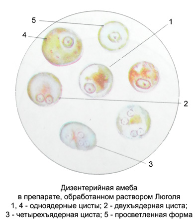
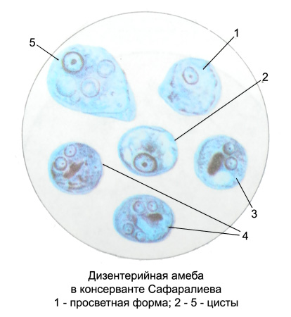
The cysts are formed from the luminal and tissue forms in the lower colon. Are found in the feces chronically ill and parasite. Is a fixed, Colorless, clear, rounded (less oval) Educational size 11 -15 M, vaginate. Sometimes cysts visible shiny sticks - chromatoid body (supply of nutrients). When processing solution Lugol clearly visible nucleus (1-4) in the form of rings and mild-defined brown spots glycogen.
Dizenteriйnaя amoeba in the luminal form a person usually lives in the upper part of the colon. However, under certain conditions it can penetrate the gut wall and become tissue pathogenic form, which leads to the development of colon inflammation and the appearance of ulcers.
Part of the amoeba with the contents of the colon fall into the rectum, where either die, or turn into cysts, are released in the feces to the external environment, which are stored for a long time. For human pathogens quad cysts (the most mature). Once in the mouth, they penetrate into the digestive canal, where their shell dissolves, and each core is halved. The result is an eight amoeba, from which then occur eight subsidiaries.
When colon tissue ulceration amoeba may enter the blood to the liver, lungs, brain and other organs and tissues, causing abscesses.
For dysentery amoeba morphologically very close intestinal amoeba - Entamoeba coli (20-40 M), which occurs in the vegetative form and in the form of cysts. In the cytoplasm of the red blood cells of its absence, but can be detected microorganisms, mushrooms, food particles. In living amoebae clearly visible nucleus. Smaller wide pseudopodia sometimes occur simultaneously in several places. The movement resembles intestinal amoeba headway. Large cysts, mostly eight- (sometimes 16- and 32-core), core have a view of the Rings. Young cysts contain one or two, less four cores. For human intestinal amoeba Nonpathogenic, common carrier (20-30 %).
What is unclear pathogenicity Entamoeba gingivalis, Dientamoeba fragile; not pathogenic to humans Endolimax nana (carriage in 15-20% of cases), Jodamoeba butcles and Entamoeba Hartmanis (carriage 5-10 % cases).
