Cellular elements in the discharge from the nipple of the breast: analysis, research to diagnose disease

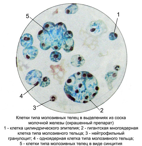
Discharge from the nipple of the breast can be observed in various diseases. Cellular composition of different secretions, most common cell type cells colostric (frothy, ksantomnye, macrophage-lipofagi). Their number may vary. Q. The origin of these cells is currently not fully resolved, assumed their derivatives as a stray elements, and epithelium. These cells are very similar to Molo- sive calves colostrum lactating women. Therefore they were called cell type cells colostric. This name is also due to the fact, that underlying pathological processes in the mammary gland disorder lie dishormonal, which may affect the secretory function of the mammary gland and invoke processes, similar to lactation.
The native preparations, these cells form round, different sizes (from 8 to 80 m), densely filled with fat droplets. The core of the large number of fat droplets seen not. In preparations, stained by Pappenheim, these bright cells, with abundant, painted in light blue tones, inhomogeneous (fine, foam or voids in place drops of fat) cytoplasm.

The kernels are small, the same magnitude, rounded, located centrally or eccentrically. The contours of the nuclei clear, smooth. The chromatin in the form of grains or clumps distributed evenly and intensely colored in purple tone. Perhaps the presence of pyknotic nuclei. The nucleoli are almost never found. The most common cell type colostric mononuclear cells, but may be multinucleated cells unlike colostrum. Arranged in isolation cells, groups, as grozdevidnyh formations, and syncytium with more cores. A significant number of these cells in the discharge from the nipple of the breast is most often seen with fibrocystic breast; but can also occur in other diseases, particularly in cancer. Therefore, only by the presence of cells such as cells in colostric discharge from the nipple of the breast is not safe to judge the nature of the pathological process.
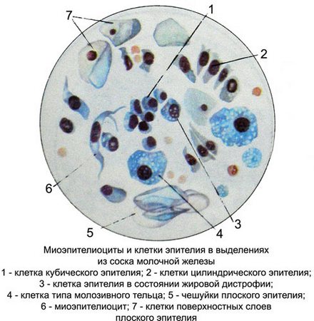
Flat epithelial cells found in the discharge from the nipple of the breast often enough, especially in those cases, When the capture tool material or a glass slide touch the nipple, and when the discharge without pretreatment take teat skin isotonic sodium chloride solution.
Stellate myoepitheliocytes in nipple discharge is rarely found. It is a medium-sized cells, elongated or fusiform, resemble fibroblasts. They may also have mnogootrostchatuyu and star-shaped and small round or oval nuclei, located in the cell center. The contours of the nuclei smooth and clear, chromatin looks nezhnopetlis- that mesh or smaller grains, well perceives coloring. The nucleoli are not found. The cytoplasm is slightly fibrous, painted in light colors, sometimes marked zone around the nucleus of enlightenment.

It is believed, that stellate myoepitheliocytes fall into isolation with ottorgshimsya proliferating epithelium, t. it is. in violation of the integrity of the epithelial lining of the milk ducts.
Cages, lining the milk ducts and lactiferous sinuses, often small or medium-sized, round or oval (Cubic or cylindrical) epithelial cells with eccentrically located nucleus and a small rim of basophilic, intensely colored or light blue cytoplasm. Perhaps the presence of cells in a state of fatty degeneration and vacuolization.
Red blood cells usually found in the discharge from the nipple amber yellow, bloody and brownish. The appearance of red blood cells may be due to injury by careless squeezing secretions or cause rejection of large areas of proliferating epithelial. In case of delay streamed blood in the lumen of the milk ducts or cystic cavities erythrocytes change, and it gives secretions brownish tint.
In describing the cytology drug should specify the number of red blood cells (a large number of, many, a moderate amount, little, single) and their character (modified or unmodified).
Neutrophilic granulocytes. When inflammation in the mammary gland number of these cells increases and they can constitute the majority of all the cells in the preparation. In the study of purulent discharge neutrophilic granulocytes densely cover the entire field of view. In such cases, they may degenerative change and decay, which is accompanied by the appearance in the preparation of cell detritus, as well as nuclear and cytoplasm ethical scraps. Among the whole cells and their fragments are often noted by many strands of fibrin.
Along with neutrophilic granulocytes are found in the product macrophages and gistiotsitы, which is characteristic of the inflammatory process. Purulent discharge from the nipple of the breast must be stained with Ziehl-Nelsenu for the detection of Mycobacterium tuberculosis and Gram to detect drusen actinomycetes.
Macrophages morphologically indistinguishable from cells such as cells colostric. They are characterized by the presence of cytoplasmic inclusions, that are clearly distinguishable in the native, and stained preparations. In inflammatory processes macrophages are identified along with neutrophilic granulocytes. Detection in spotting macrophages, containing hemosiderin or fragments erythrocytes, It indicates the presence of cavities, filled with blood. Most often this occurs when benign dysplasia (mastitis) with intraductal epithelial proliferation and fomirovaniem papillae.
Gistiocitы - A variety of largest, shape and color of tissue cells. Their diameter ranges from 10-20 mm. The kernel oval or bean-shaped, with delicate chromatin structure, located in the center or eccentrically. At the core there is a 1-2 nucleoli, which can not be viewed due to degenerative changes in the cell. Basophilic cytoplasm, painted in different shades of blue tones, often vacuolated.
Fibrocytes and fibroblasts - Elongated cells with oval or rod-shaped hyperchromatic nucleus. Basophilic cytoplasm, often located only poles of the cell. Fibroblasts unlike fibrocytes have a young nucleus with nucleoli and more intensely colored basophilic cytoplasm.
Gistiocitы, fibroblasts and fibrocytes are most often found in inflammatory processes, and during the attenuation process and organization hearth degradation may even dominate the formulation.
Giant multinucleated cells. The discharge from the nipple may be found giant cells colostric cells, Pirogov-cells and Langhans cells and foreign bodies.
