Fabric classification
Cloth --eto phylogenetically developed system of cells and non-cellular structures, having the common structure and specialized to perform specific functions. Depending on this distinguished epithelial, derived mesenchymal, muscle and nerve tissue.
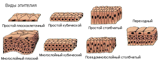
Epithelial tissue morphologically characterized by close association of cells in layers. Epithelium and mezotelïy (kind of epithelium) lining the surface of body, serous membranes, the inner surface of hollow organs (alimentary canal, bladder, etc.. d.) and form the majority of the glands.
There are covering and glandular epithelium
Pokrovnıy epithelium It refers to the border, because it is located on the border of the internal and external environments, and through the exchange of substances (absorption and excretion). It also protects the underlying tissues from chemical, mechanical and other external influences.
Glandular epithelium It has secretory function, t. it is. the ability to synthesize and secrete substances Secrets, has a specific effect on the processes, occurring in the body.
The epithelium is located at the basement membrane, beneath which lies a loose fibrous tissue. Depending on the ratio of cells to the basement membrane differentiate single and multilayer epithelium.

Epithelial, all cells which are connected to the basement membrane, It called single-layer.
In stratified epithelium basement membrane bound only the bottom layer of cells.
There are one- and Multi odnosloynıy epithelium. For single-row isomorphic epithelial cells characterized by the same shape with nuclei, lying flush (par), and for multi-row, or anizomorfnogo - cells with nuclei of various shapes, lying on different levels and in several rows,.
Mnogosloynıy epithelium, wherein the cells of the upper layer are converted into horny scales, called multi-layered stratum, and in the absence of keratinization - multilayer neorogovevayuschy.
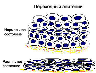
A special form of stratified epithelium is the transition, characterized, that its appearance changes depending on the underlying tissue stretching (the wall of the renal pelvis, ureters, bladder and others.).

Through a single-layer single-row epithelium is an exchange of substances between the organism and the environment. For Example, monolayer epithelium of the alimentary canal allows the absorption of nutrients in the blood and lymph. Multilayer (epithelium Kozhim), and monolayer epithelium (bronchi) It performs primarily defensive function.
Cloth, develops from the mesenchyme
Blood, lymph and connective tissue develop from a bud of tissue - mesenchyme, therefore join the group of musculoskeletal tissue trophic.
Hemolymph - Fabric, consisting of the extracellular liquid substance freely suspended therein cells. Blood and lymph perform trophic function, carry oxygen and various substances from one organ to another, humoral providing connection of all organs and tissues.
Conjunctive tissue divided into the actual coupling, cartilage and bone. It is characterized by the presence of large amounts of fibrous intercellular substance. Connective tissue performs trophic, plastic, protecting and supporting functions.
Muscle tissue
There are neischerchennuyu (smooth) muscle, consisting of elongated cells, and striated (striated) , formed muscle fibers, have sim- plastic structure. Neischerchennaya muscle tissue develops from the mesenchyme, and striated - from the mesoderm.
Nervous tissue
Nervous tissue It consists of nerve neuron kletok-, the main function of which is the perception and conduction of excitation, and glia, organically linked to nerve cells and performing the trophic, mechanical and safety features. The rudiment of the nervous system at an early stage of embryo development stands apart from the ectoderm, except microglia, derived from the mesenchyme.
The development of tissue – norm and pathology
With fabrics such concepts are related, how proliferation, giperplaziya, metaplasia, Dysplasia, anaplasia and regeneration.
Proliferation - All kinds of cell proliferation and intracellular structures in normal and pathological conditions. It lies at the heart of the growth and differentiation of tissues, It provides a continuous renewal of cells and intracellular structures, and reparation processes. Proliferation kletok, lost the ability to differentiate, It leads to tumor formation. Proliferation is the basis of metaplasia. Different tissues have different abilities to proliferate. High proliferative capacity different hematopoietic, connective, bone tissue, epidermis, mucosal epithelium, moderate - skeletal muscle, pancreatic epithelium, salivary glands, etc.. Low proliferative ability, or lack of it is typical for the central nervous system tissue and myocardium. When the function of the tissue damage is reduced by intracellular proliferation. The proliferation of intracellular structures leads to an increase in cell volume, their hypertrophy. Hypertrophy of the body as a whole may occur due to both cellular, and intracellular proliferation.
Giperplaziya - Increasing the number of cells by their excess tumors. Carried out by means of direct (mitosis) and indirect delenyya (amitoza).
By increasing the number of cell organelles (riʙosom, mitochondria, the endoplasmic reticulum and others.) talk about the intracellular hyperplasia. Similar changes are observed in hypertrophy. Hyperplasia is a part of proliferation, as the latter covers all types of cell proliferation in normal and pathological conditions. Hyperplasia develops as a result of a variety of influences, stimulating cell proliferation, it is the result of overproduction of cellular elements. In addition to increasing the number of cells characterized by hyperplasia and some of its qualitative changes. Cells are larger than the original, uniformly increasing their nucleus and cytoplasm number, whereby nucleocytoplasmic ratio is not changed. There may be nucleoli. Cell hyperplasia with atypia is regarded as dysplasia.
Metaplasia - Stable transformation of one fabric type to another by changing its morphology and function. Metaplasia can be direct - to change the type of fabric without increasing the number of cellular elements (converting the actual connective tissue into bone without osteogenic elements) and indirect (tumor), which is characterized by cell proliferation and differentiation. Metaplasia can occur on the basis of chronic inflammation, lack of retinol in the body (vitamin A), of hormonal status, etc..
The most common epithelial metaplasia, eg metaplasia of columnar epithelium in flat (in the bronchi, salivary glands and sebaceous, bile ducts, intestines and other organs, having glandular epithelium) or intestinal metaplasia (эnterolizatsiya) epithelium of the mucous membrane of the stomach gastritis.
The transitional epithelium of the urinary bladder in chronic inflammation may metaplazirovatsya and flat, and glandular. Squamous epithelium of the oral mucosa in a flat stratum metaplaziruetsya. Conclusive evidence of the transformation of connective tissue into the epithelial not available.
Dysplasia - Abnormal development of organs and tissues during embryogenesis and postnatal (postpartum) period, when the effect of intrauterine factors manifested after birth, Even in an adult.
In oncology, the term "dysplasia" is used to determine the precancerous tissue, involving a violation of regeneration, which proceeds as hyperplasia (with excessive formation of cells) and always with signs of atypia.
Depending on the degree of cell atypia are three degrees of dysplasia:
- Easy;
- Moderate;
- Heavy.
Mild dysplasia It is characterized by the emergence of dual-core in single cells while preserving normal cells in the rest of the nuclear-cytoplasmic ratio. In some cells there may be signs of degeneration (vacuolar, fatty and other.).
In moderate dysplasia in single cells marked increase in the appearance of nuclei and nucleoli in them.
Severe dysplasia characterized polymorphism cells, annzotsytozom, increasing cores, granular chromatin structure therein, the advent of multi-core cell. The nuclei are found nucleoli. The nuclear-cytoplasmic ratio changes in favor of the core. In the cells appear more pronounced degenerative changes. Location chaotic cells. Cytologic dysplasia is difficult to distinguish this from intraepithelial cancer. In cases of severe dysplasia, abnormal cells are not many, as in carcinoma in situ (preinvasive cancer - a malignant tumor in the early stages of development).
According to some researchers dysplasia is mild to moderate and rarely progresses 20-50 % cases regress.
With regard to severe dysplasia, there are different points of view: Some scientists believe, It is possible that the reverse development and transformation into cancer; according to others, severe dysplasia is an irreversible condition, which necessarily becomes cancer. Phenomena dysplasia can also occur with indirect metaplasia.
Anaplasia - Persistent violation of maturation of cancer cells to change their morphology and biological properties. There are biological, biochemical and morphological anaplasia.
Biological anaplasia characterized by loss of cell functions, except for the function of reproduction.
Biochemical anaplasia cells manifested loss of the enzyme systems, characteristic of stem cells.
For anaplasia characteristic morphological changes in intracellular structures, and the shape and size of cells.
