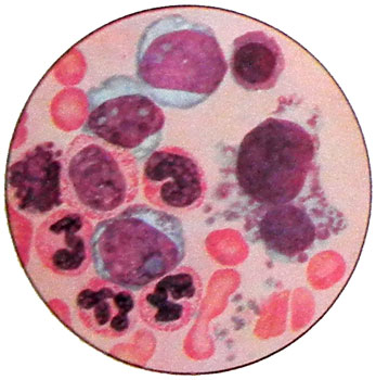
Chronic myelogenous leukemia
Chronic myelogenous leukemia - a tumor, arising from early progenitor cells myelopoiesis, differentiating into mature forms; cell substrate it up predominantly neutrophilic granulocytes. The disease is naturally two stages: detailed benign (monoklonovuyu) and terminal malignant (polyclonal). Chronic myelogenous leukemia occurs most often in adults aged 30-70 years, and men often, than among women.
In 86-88 % cases of chronic myeloid leukemia in virtually all bone marrow cells, except lymphocytes - granulocytes, monocytes, erythrokaryocytes and megakaryocytes - revealed Ph'-chromosome (Philadelphia), opened in 1960 g.
Although, seemingly, leukemia is all three germ bone marrow, unlimited growth is inherent in the expanded stage, usually, Only one germ - granulocytic. Less common concerns and increased production of megakaryocytes, and platelets.
Stages of chronic myeloid leukemia
True initial stage chronic myeloid leukemia, when only a small portion of bone marrow cells contain Ph'-chromosome, and make up a significant percentage of cells without Ph'-chromosome, detected extremely rare.
Usually, disease is diagnosed in step total generalization tumors in the bone marrow with extensive proliferation of tumor cells in the spleen, and often in the liver, t. it is. in advanced stage of chronic myeloid leukemia.
Pathology developed stage chronic myeloid leukemia It is characterized by the growth in the bone marrow myeloid tissue, almost complete displacement of fat in the flat bones, the advent of bone marrow hematopoiesis in long bones (epiphysis, diafiz), proliferation of myeloid tissue in the spleen, liver. At the same time there is a proliferation of trehrostkovaya, predominates granulocyte germ.
The bone marrow is usually somewhat increased content of megakaryocytes, These cells are found in spleen and. Lymph nodes in the advanced stage of the disease is usually not affected the process of leukemia. In some cases, the bone marrow can develop myelofibrosis, but it happens more often after prolonged therapy mielosanom. Women mielosanom therapy rather quickly leads to amenorrhea, and therefore marked hypoplasia of the uterus, atrophy of the mucous membrane, hypoplasia of the ovaries. The skin of patients, especially women, taking long mielosan, It has a grayish-brown hue - expressed melasma. Chance of pulmonary fibrosis. The infiltration of the liver tumor cells in most cases pronounced.
The pathogenesis of the advanced stage of chronic myeloid leukemia, manifested primarily in the development of asthenic syndrome (weakness, fatigue, etc.. P.), due to increased cell disintegration, which in some cases may be accompanied by an increase in uric acid in the blood (hyperuricemia), urine (giperurikurija), the appearance of kidney stones, but it may be due to the peculiarities of production and granulocytes, eg gistaminemiey. At very high leukocytosis, reaching 500 T in 1 l and more, may develop multiple circulatory disorders, primarily in the brain, in connection with the formation of leukocyte stasis (in the gut such stasis may be complicated by bleeding).
In recent decades, due to the development of chemotherapy similar levels of white blood cells in chronic myeloid leukemia almost never occur. In the advanced stage of severe anemia is usually not observed, although in some cases the level of red blood cells may be somewhat lower limit of normal. Thrombocytopenia occurs not often. Genesis tsitopenichesky such situations has not been elucidated. Least likely cytolysis, since reticulocytosis, increased number of bone marrow erythrokaryocytes, increased bilirubin in blood or urine hemosiderin are not observed.
Advanced stages of chronic myeloid leukemia is characterized monoclonal myeloid cells, the practical elements of the displacement of normal hematopoiesis: cells Ph'-chromosome in the bone marrow are about 98-100 %. However, a few years later when the chromosomal analysis can detect when a single cell with an abnormal number of chromosomes (often - giperdiploidnyh), which can be a source of new subclones, Beginners terminal stage of the process. All new versions karyological cell Ph'-chromosome is always preserved.
Clinical manifestations of chronic myeloid leukemia
Clinical manifestations of the initial stage of the disease can not be determined. The first symptom is a leukocytosis with a shift to myelocytes and promyelocytes in the background of the normal state of health of the patient. With increasing leukocytosis occur sweating, weakness, fatigue. These symptoms usually appear already at leukocytosis, in excess of 20-30 g 1 l. In some patients, and the level of white blood cells over 200 T in 1 l What- or no discomfort, but it is rare. Sometimes, the first symptoms of disease severity are, slight pain in the left upper quadrant due to an enlarged spleen, develops 85 % patients.
In the absence of cytostatic therapy, the disease gradually progresses: narastaet leukocytosis, increased spleen and liver, deteriorating health (weakness, fatiguability, Sweating).
Blood picture of chronic myeloid leukemia
Picture of blood in the advanced stage of chronic myeloid leukemia is characterized by neutrophilic leukocytosis with a shift to myelocytes, promyelocytes and myeloblasts even single.

Red blood at the beginning of the disease does not change significantly, sometimes marked presence of single erythrokaryocytes. The platelet count in the individual cases can be reduced, but more often it is normal. The 20- 30 % cases from the beginning of the disease is marked thrombocytosis, which can reach high numbers: 1,5-2.0 T 1 l and more. In the absence of treatment is steadily increasing leukocytosis, The number of platelets or remained stable, or slowly increasing.
In the advanced stage of chronic myeloid leukemia bone marrow is very rich in cellular elements. In trepanate it noted the almost complete replacement of fat primarily granulocytic cells. At high thrombocytosis observed a large number of megakaryocytes. In a smear of bone marrow granulocytes predominate, whereby the ratio reaches leykoeritroidnoe 10:1, 20:1 and more. The morphology of the blood cells and bone marrow were not significantly different from the norm. Only in granulocytes usually noticeable decrease in grain.
Pathological granulocyte maturation of the germ in the advanced stage of chronic myeloid leukemia seen changes in the content and specific azurophilic granules, sometimes lack, low myeloperoxidase in promyelocytes, myelocytes, mature neutrophils, in some cases in the presence of enzymes along with myelocytes granulocytic monocytic series of several enzymes. Reduction of mature neutrophils alkaline phosphatase is a characteristic feature of the advanced stage of chronic myeloid leukemia. Alkaline phosphatase in cells of the patient may be absent.
One of the characteristics of chronic myeloid leukemia is a high content of cobalamin (Vitamin B12) in serum and its high capacity to bind them. Elevated levels of serum cobalamin due to the high level of transcobalamin I - one of the transport proteins, secreted by cells of granulocytic series.
In addition to leukogram rejuvenation of granulocytes can be observed increase in the percentage of basophils and eosinophils, less of both at the same time (basophilic-eosinophilic Association). Besides, in the blood can be detected and isolated blasts erythrokaryocytes.
The punctate enlarged spleen developed stage reveals the predominance of myeloid cells.
Gradually, the process progresses, resulting in a slow, but steady increase in the spleen, gradual increase leukocytosis, to reduce that there is a need to increase the dose, and in some mielosana lower rates of red blood and platelet counts.
At some, advance unpredictable, the process proceeds from step monotonically growing benign tumor malignant polyclonal, t. it is. disease in the terminal stage proceeds. Clinically it is manifested by a sudden change of the whole picture of the disease: a rapid increase in the spleen, her appearance in heart attacks, any increase for no apparent reason in body temperature, or severe pain in the bones, or development of dense pockets of sarcomatous growth in the skin, lymph nodes, etc.. P. All of these new manifestations of the disease are associated with the occurrence in the main tumor clone new mutant subclones, lose the ability to differentiate, but having the ability to proliferate, displacing the initial differentiating cell clone. Occasionally manifestations of the disease start with end-stage.
There is no single symptom, mandatory for the terminally ill, as well as a combination of mandatory signs, not available. The terminal stage is characterized by a change in the quality characteristics of leukemia. Here are pronounced signs of tumor progression, which was not there before. Usually comes thrombocytopenia, in some cases there is a profound leukopenia in the absence of blasts in the blood.
The most common kind of deformation preceded blastosis leukogram - reducing the percentage of segmented and stab neutrophils with an increasing number of myelocytes, about- mielotsitov, blast cells, representing in aggregate still few percent.
Blast crisis of chronic myeloid leukemia
Hematologic changes in end-stage chronic myeloid leukemia often characterized by blast crisis - a rapid increase in the content of blasts in the bone marrow and blood. Of a certain level of blasts, corresponding to the terminal stage, does not exist. In very rare cases and in advanced stage of leukemia blasts can reach 5-10 and even 15 % and stay at that level for a number of years without any signs of change in the nature of the pathological process. When the blast Stroke increases and the number of blasts, Besides, changes their morphology: there are atypical forms with wide cytoplasm, irregular contours of the nucleus and cytoplasm.
The morphology of the blast crisis is very varied. It can be mainly myeloid, or myelomonoblastic, or monoblastic, or erythroblastic (picture of acute erythremic myelosis), or megacaryoblastic.
Cytochemical analysis, usually, allows the identification of blast cells, which represented a crisis. They may contain enzymes early stages of maturation of granulocyte series (hloratsetatesterazu more often, than peroxidase), often both enzymes monocytic series, indicating that supplies of blast cells to early seed colony cell culture (CFU-GM) before its differentiation into myeloblasts and monoblasty.
Эritroblasticheskaya nature blastnogo crisis It can be confirmed morphologically, and using a combination of a cytogenetic analysis cytochemical (dyed sideroblasts).
At the blast cells, with clear signs of cytochemical founders of a series, in the blood and bone marrow are blasts and non-differentiable elements. Apart from those cases, when the blast crisis goes through several stages of morphological, dedifferentsiruyas of morphologically undifferentiated myeloid, there are also such, when it is already at the first appearance in the terminal stage of blast cells are morphologically unidentifiable and cytochemically. The emergence of just such cells was the basis for the interpretation of the relevant cases, blast crisis as a lymphoblastoid. Application of the method of cultivation of these leukemic cells (unidentified blasts) Agar diffusion chamber and allowed to establish, that a large percentage of cases they grow in agar and respond to colony stimulating factor as myeloid cells, or even to a more differentiated mature cells of granulocytic series.
In some instances, undifferentiated blastic using only electron microscopy blast cells were detected simultaneously and myeloperoxidase glycogen depot, which in the light microscope under cytochemical analysis it looked like glycogen granules lymphoblasts.
In one embodiment, the end-stage chronic myeloid leukemia, a sharp increase in the number of basophils. Sometimes they are presented mostly mature forms, sometimes - Young, up to grit blast forms, like basophils.
Monocyte crisis in chronic myeloid leukemia
Pretty rare option is monotsytarnыy crisis - The emergence and growth of the number of mature, Young and atypical monocytes in the blood and bone marrow.
In connection with the violation of the bone marrow barrier characteristic feature of end-stage it is to detect in the blood fragments of the nuclei of megakaryocytes (in advanced stage, they appear very rarely, only at a very high level of platelets), and erythrokaryocytes (mielemiya). The most important element of end-stage regardless of the morphological picture of depression is normal hematopoiesis. It granulozitopenia, thrombocytopenia and anemia are the immediate causes of complication.
In some cases, the beginning of the terminal phase is characterized by a rapid increase in the spleen. With its high percentage of punctures found blasts. In the presence of dissociation between blastosis in spleen and bone marrow, wherein the content of blasts may be normal, there are grounds for splenectomy. The blood can be detected with high blastosis by entering the cells from the spleen. Often a symptom of end-stage liver is to increase the development of myeloid tissue in it.
Occurrence leykemidov in chronic myeloid leukemia
One of the manifestations of end-stage - the emergence of the skin leykemidov. Usually, they are represented by blast cells, However, there are (rarely) leykemidy from more mature granulocytes - promyelocytes, mielotsitov, until segmented.
Leykemidy look like slightly raised above the surface of the skin brown spots or pink (leykemidy of mature cells do not change color). The consistency of thick, They are painless. The appearance reflects the emergence of a new leykemidov subclone cells, Deprived of their tissue specificity. However zrelokletochnye leykemidy may in the long term is not transformed into a blast, This transformation is generally not required. Appearing in one place, leykemidy blast tend to metastasize to other parts of the skin, and then in the internal organs and systems.
Recently, in connection with the extension of life of patients in the terminal stage of a relatively common neuroleukemia, clinically no different from those in acute leukemia.
Growth of cells in the lymph nodes
Another focus of the growth of cells are lymph nodes, where developing solid tumors such as sarcomas, cells (blasts) which found Ph'-chromosome. The emergence of sarcomatous lymph node in chronic mielolenkoze always mean offensive end-stage. Pockets of growth of sarcoma can occur in any organ ", disrupting its function, and bone.
If the terminal stage begins with the proliferation of bone marrow blasts off, in some cases it is possible to suppress the process, which for some time local, or chemotherapy, or radiotherapy, or splenectomy (in isolated localization of blasts in the spleen) and receive remission, sometimes quite long. However, this does not mean, that the progression of the tumor process in chronic myeloid leukemia is associated with the spleen and prior removal of the body can delay the onset of end-stage.
The most important and earliest sign of approaching the terminal phase and blast crisis is the development of refractory to mielosanu. Often, use of this drug in the early stage of the terminal tends to reduce the number of leukocytes, However, the size of the spleen or liver is not reduced or even increased. This kind of partial refractory mielosanu sometimes develops until the clearest signs blast crisis.
IN 90 % cases of end-stage chronic myeloid leukemia found aneuploidija. Dominated giperdiploidnye clones.
Chronic myeloid leukemia in children
Chronic myelogenous leukemia in children are divided into two forms - infantile, that dominates under 3 years, and juvenile, is more common after 5 years. The frequency of chronic myeloid leukemia in children is 1.5-3 % all leukemias.
The infantile form of chronic myeloid leukemia
The infantile form of chronic myeloid leukemia differs from chronic myeloid leukemia in adults a number of features. The main one is the lack of Ph'-chromosome, although there may be other non-specific chromosomal abnormalities.
The red blood cells significantly increased the content of fetal hemoglobin: the level reached 100 % (at a rate of less than 2 %). Obviously, Fetal hemoglobin is produced in this case a number of leukemic cells red.
Picture of blood at the infantile form of chronic myeloid leukemia It is characterized by a tendency to thrombocytopenia in the advanced stage of the disease, often observed monocytosis and eritrokariotsitoz, increase in the percentage of immature forms. The serum and urine of patients with significantly increased content of lysozyme.
Clinically, infantile form of lymphadenopathy seen a moderate increase in spleen, often the appearance of rashes on the face, susceptibility to infections during this form of chronic myeloid leukemia adverse, the average life expectancy does not exceed 8 months.
The juvenile form of chronic myeloid leukemia
The juvenile form is characterized by Ph'-chromosomes in myeloid cells. This form of the disease is not very different from chronic myeloid leukemia in adults, although in the advanced stage of her children are often found lymphadenopathy, increase not only the spleen, and liver.
Chronic myelogenous leukemia without Ph'- chromosome in children and 9-15 % Adult. Different unfavorable course and short life expectancy of patients.
Especially It has a poor prognosis with chronic myelogenous leukemia Ph'- chromosome, flowing a tendency to thrombocytopenia already advanced stage or high myelocytes and even blast cells in the peripheral blood (a moderate increase in the level of white blood cells).
In a particular embodiment should be allocated chronic myeloid leukemia in patients over the age of 60 years. It is often characterized by the slow development of the process and the duration of life of patients.
