Glomerulonephritis – state and urinalysis
Currently, it is generally accepted, that glomerulonephritis (GN) It is an immune-inflammatory disease.
Acute glomerulonephritis
The most common occurrence of acute glomerulonephritis associated with streptococcal infection (pharyngitis, tonsillitis, diseases of the skin and so on.). Most nephrogenic is β-hemolytic streptococcus (Types 12, 49) Group A. There are cases of acute glomerulonephritis in patients with diseases of staphylococcal etiology, particularly acute staphylococcal endocarditis. It is also possible immediately after the development of glomerulonephritis lobar pneumonia, typhoid fever, malaria, infective hepatitis, peel, chicken pox, etc.. d. The disease can also occur due to the strong cooling, especially when exposed to cold wet.
The main clinical symptoms are swelling, hypertension and hematuria.
Swelling - The earliest and constant symptom of acute glomerulonephritis. Their pathogenesis is still not fully understood, but it is believed, that the most important role is played by renal dysfunction, resulting in a delay of sodium chloride and water in the body. In acute glomerulonephritis there is disruption and filtering, and reabsorption, which ultimately leads to the appearance of edemas. Filtering it reduces (retained water and sodium), reabsorption of sodium, and with it, and water, increases. Thus, there is a significant delay of water and sodium in not only blood, and tissue; this contributes aldoste- Ron, which retains the body and sodium, Consequently, water (aldosteronism) and acute glomerulonephritis released in an increased amount.
Hypertension in acute glomerulonephritis due to the fact, that this disease in the body, one side, are formed in increased amounts and the renin-angiotensin-, with another - increased fluid content. The role of the complex of the renin - angiotensin system in the development of hypertension in this disease is confirmed by the work of several researchers, which describes the juxtaglomerular complex hyperplasia with acute glomerulonephritis, flowing with high blood pressure. In the development of hypertension in acute glomerulonephritis is also important, and increased secretion of aldosterone (secondary aldosteronism), contributes to the accumulation of sodium in the walls of arterioles, which leads to their swelling, toning and hypertensive reactions. Thus, in acute glomerulonephritis secondary aldosteronism plays a role in the development of edema, and Hypertension.
Pathological changes in the kidneys in acute glomerulonephritis are caused by the deposition in the glomerular capillaries heterologous immune complexes. According to the morphological picture of acute glomerulonephritis refers to a proliferative form of endokapillyarnoy, in which there is a sequence of various phases of development: exudative, exudative-proliferative, proliferative phase, and residual effects.
Microscopic examination drugs found a picture of diffuse kapillyarita. All pochechnыe balls uvelychenы. The endothelium of the capillaries and mezangiotsity (mesangial cells) most are in the state of active proliferation and swelling. Mesangial infiltrated by polymorphonuclear leukocytes. Severe congestion of the capillary network and presence in the oral capsule glomerular hemorrhagic exudate possible to identify hemorrhagic form of acute glomerulonephritis.
The predominance of white blood cells indicates the exudative phase (form), a combination of cell proliferation and glomerular leukocyte infiltration seen as exudative-proliferative phase, and the predominance of cell proliferation - as the proliferative phase (shape) acute glomerulonephritis.
According to electron microscopic study, in acute glomerulonephritis occur thickening and swelling of the capillary basement membrane, thinning, splitting, cavities and fractures.
Changes in the tubules of nephrons the first time or there is no hyaline droplet, less vacuolar degeneration of the epithelium of proximal tubules. The lumina of the tubules found erythrocytes, cylinders, sometimes white blood cells.
When the disease by reducing the filter and improve kidney function observed reabsorbtsionnoy oligurija. The oliguric phase relative density of urine is 1,022-1,032, which should be considered in the diagnosis of chronic nephritis.
In acute glomerulonephritis observed breaks capillaries, leading to urinary excretion of protein fractions and erythrocytes, and it can be combined with a decrease in filtration. High concentrations of protein in urine at an acute nephritis depends on the reabsorption of water. Standing is a sign of acute glomerulonephritis hematuria. It is observed in the majority of patients with acute nephritis, but the extent of it is different from the gross hematuria, (urine color of meat slops) to mykrohematuryy (10-15 erythrocytes in the field of view). Hematuria can not be explained only by increasing the permeability of the glomerular filter. Histologically hematuric glomerulonephritis detected breaks capillaries and blood clots in the glomerular capsule, with the urine can contain little protein and a lot of red blood cells. The amount of protein in urine varies from 2-3 to 20-30 g / l. The reaction is weakly acid urine, precipitate it in some cases brown, loose, which affects the color and opacity of urine.
Microscopic examination of the urine showed normal white blood cell count, but it is possible to increase to 20-30 in their field of view. Red blood cells are found in varying amounts, often leached, sometimes fragmented; can be observed and unaltered, especially in severe hematuria.
Renal epithelial cells are found in different quantities, in severe cases - in a state of fatty degeneration.
Cylinders (hyaline, grainy, epithelial, buropigmentirovannye, blood) are found in varying amounts, fibrin burookrashennыy. Observed granular breakdown of hemoglobin and uric acid crystals.
Classic for acute glomerulonephritis recently in adults is rare. Often there is erased clinical picture, Only limited urinary syndrome, often faint.
Acute glomerulonephritis may result in spontaneous recovery, or go to the subacute. The latent form of acute glomerulonephritis sometimes becomes chronic is not- froticheskiy glomerulonephritis. If glomerulonephritis does not pass within a year, it should be considered a chronic.
Subacute (rapidly progressive) glomerulonephritis
In this form of the disease is detected morphologically extracapillary proliferative processes. From the point of view of pathogenetic identify several forms of rapidly progressive glomerulonephritis:
- idiopathic;
- pulmonary-renal syndrome, hereditary (Goodpasture's syndrome) - Disease, due to the appearance of anti-glomerular basement membrane antigen;
- immune-complex, etc..
Features of change of the glomerular renal corpuscles in the subacute glomerulonephritis is necrosis of the capillary walls and gaps, whereby blood is poured into the cavity of the capsule and the glomerulus falls fibrin. Proliferation of glomerular epithelial capsule leads to the formation of peculiar crescents, ohvatыvayuschyh and szhymayuschyh pochechnыe balls. Epithelial crescent gradually converted into fibrous, and then creating sclerosis and gialiniziruyutsya.
The tubules of nephrons observed hyaline droplet and vacuolar degeneration of epithelial cells. The disease leads to rapidly progressive destruction of nephrons, death occurs from renal failure.
Clinically, the disease begins as a typical, less as a latent form of acute diffuse glomerulonephritis: severe swelling until anasarca, high blood pressure, severe retinopathy with retinal detachment, hypoproteinemia (to 31,6 g / l), hypercholesterolemia (to 33,8 mmol / l). There is a progressive decline in kidney filtration function, and from the very first weeks of the disease may increase azotemia, that leads to the development of anemia.
This disease is characterized by oliguria, where the first observed high relative density of urine, then decline rapidly, despite the expressed oliguria.
Proteinuria dostigaet 102,8 g / l. There hematuria (erythrocytes unchanged, leached and fragmented). Epithelial cells of the kidneys partly steatosis and vacuolation. There hyaline, grainy, epithelial, buropigmentirovannye, blood, hyaline droplets and other cylinders can detect burookrashenny fibrin and grain hemosiderin.
Chronic glomerulonephritis
Chronic glomerulonephritis is often the result of acute uncured. However, he often develops without prior acute attacks, t. it is. as the primary chronic glomerulonephritis. The etiology and pathogenesis of the same, as in acute nephritis.
In chronic glomerulonephritis primarily affects glomerular kidney cells. The defeat of this character is intracapillary. First, the kidneys are not changed, in the future with the development of fibrotic process, they shrink, significantly reduce (secondary contracted kidney). Microscopic examination of observed changes in the glomerular capillary wall thickening in the form (proliferation, gialinoz, proliferation of connective tissue), leading to narrowing of the lumen of the capillaries, and even its complete closure. The basal membrane thickens, then it appear fibrotic changes. The capsule as glomerular proliferative changes occur, resulting in narrowing the lumen of the capsule and it turns into a narrow gap. The tubules of nephrons expressed degenerative changes (zernistaя, and further adipose and hyaline- kapelynaya dystrophy). With the progression of the process there is a complete cessation of glomerular renal function and death of cells corresponding to tubules of nephrons. Thus, some nephrons completely fail.
The main clinical symptoms of the disease – swelling, hypertension, hypoproteinemia, cholesterolemia, proteinuria and gematuriya, expressed in varying degrees. Distinguish following clinical forms of the disease:
- latent;
- hematuric;
- hypertonic;
- nephrotic;
- mixed.
The most common latentnыy glomerulonephritis. It manifests itself only slightly pronounced and urinary syndrome is often a moderate increase in blood pressure. In the investigation of urine revealed mild proteinuria, mikrogematuriâ, individual hyaline and granular casts.
Gematuricheskiy glomerulonephritis rare (in 6 % cases), It characterized by constant hematuria, sometimes gross hematuria. In this form of the disease observed in urine volume, bloody or loose brownish pellet.
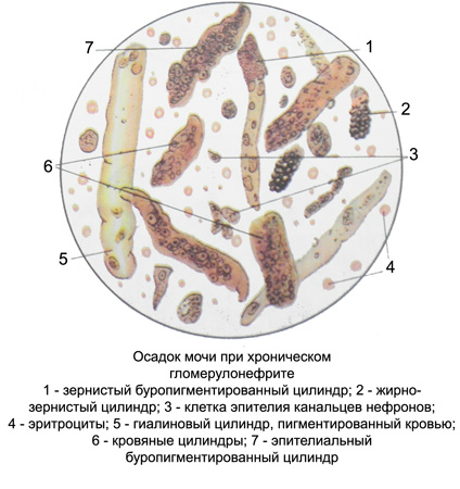
Microscopically detected at microhematuria leached and fragmented erythrocytes, in cases of gross hematuria - unchanged, leached and fragmented. Found hyaline, grainy, epithelial, buropigmentirovannye, blood, hyaline droplets, vacuolated, Sometimes fatty granular casts. Renal epithelial cells are in a state of granular and vacuolar degeneration of fat, buropigmentirovannye blood, from one to several copies per microscopic field, sometimes form small clusters. On the morphological elements of the urinary sediment found burookrashenny fibrin shreds and hemosiderin as amorphous masses.
Independent forms of chronic glomerulonephritis should be considered hematuric glomerulonephritis deposition in the glomeruli of the renal corpuscles lgA-lgA-hlomerulopatyyu (Berger's disease), observed more often in young men after a respiratory infection and often occurs with gross hematuria.
Nefroticheskiy glomerulonephritis It is characterized by severe edema,, massive proteinuria (More than 4 5 g per day), hypercholesterolemia (more specifically hyperlipidemia) and hypoproteinemia (by albumin). Blood pressure is normal or low. Urine output decreased.
In moderately progressive course of nephrotic glomerulonephritis morphologically appears as membranous or mesangioproliferative. In cases where a more rapid progression of the disease are observed mesangiocapillary glomerulonephritis. focal segmental glomerulosclerosis, or glomerulonephritis fibroplastic.
The number of leukocytes in the urine within normal, in some cases, it increases to 30-40 copies per field of view of the microscope. Within microhematuria meet and unchanged erythrocytes. Renal epithelial cells are in a state of granular and fatty degeneration. Cylinders hyaline, grainy, epithelial, buropigmentirovannye, blood, hyaline droplets, fat-grained, vacuolated and in severe cases, waxy.
Hypertensive glomerulonephritis initially, usually, It has latent within. In the urine there is a slight proteinuria and microscopic hematuria (leached erythrocytes), single cells of renal epithelium and hyaline, granular casts. Diagnosis of this form of chronic glomerulonephritis causes significant difficulties. Hypertension often benign nature. Long-term course of the disease, gradually progressing, mandatory outcome in chronic renal failure.
Mixed glomerulonephritis characterized by a combination of nephrotic syndrome and hypertension. Swelling in this form can be significant, and hypertension is somewhat less pronounced, than in hypertensive form.
Thus, changes in urine, as well as the clinical manifestations of chronic glomerulonephritis, diverse. Oliguria is not expressed, the amount of urine and its relative density is often normal. With the development of renal failure appears polyuria, and then, at the secondary contracted kidney - oliguria with gipoizostenuriey. The amount of protein in urine varies depending on the clinical disease. At a glomerulonephritis nephrotic proteinuria is more pronounced, than when hematuric. When latent glomerulonephritis protein in the urine slightly, and at secondary contracted kidney even less, indicating that the death of the nephrons.
The number of red blood cells is also different, they are predominantly leached, often subtle and fragmented, but nephrotic form of the disease may be unchanged. Degenerative changes in the cells of the renal epithelium is usually more pronounced, than in acute glomerulonephritis. It has not only hyaline, grained, epithelial, buropigmentirovannyh and blood, and hyaline droplet, fatty granular and waxy cylinders indicates the severity of the process.

There are patches of fibrin burookrashennogo. There is a granular breakdown of hemoglobin. In severe cases, the death of many nephrons amount of urine, cylinders and protein in the urine decreased. With the development of secondary contracted kidney and renal failure occur poly- and izostenuriya, low protein content in urine, in the sediment - wide cylinders, originating from excessive expansion preserved hypertrophic tubules of nephrons.
