Red blood cells - red blood cells Types – Erythrogenesis
Erythroblast
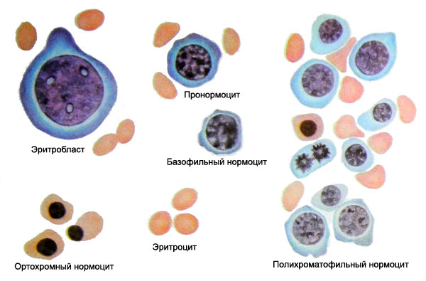
Erythroid progenitor cells is the erythroblast. He comes from a cell eritropoetinchuvstvitelnoy, which develops from a progenitor cell myelopoiesis.
Erythroblasts up diameter of 20-25 microns. The core of it is almost geometrically round shape, painted in red and purple. Compared with non-differentiable blasts can note a rough texture and a bright color core, although chromatin fibers rather thin, interweaving their uniform, nezhnosetchatoe. At the core are two - four nucleolus and more. The cytoplasm of cells with a purple tinge. Around the nucleus there is enlightenment (perinuklearnaya area), sometimes with a pink tinge. These morphological and tinctorial features make it easy to recognize erktroblast.
Pronormocit
Pronormocit (pronormoblast) Like erythroblasts characterized clearly defined round core and pronounced basophilic cytoplasm. Distinguish pronormotsit from erythroblasts can be at a rough structure of the nucleus and the absence of it nucleoli.
Normocytic
Normocytic (Loevit cell) largest non-nuclear approaches mature erythrocytes (8-12 M) with deviations in one direction or another (Micro- and macroform).
Depending on the degree of saturation of hemoglobin distinguish basophilic, polychromatic and oxyphilic (ortohromnye) normocytes. The accumulation of hemoglobin in the cytoplasm normocytes happens with the direct participation of the nucleus. This is evidenced by the appearance of his first around the nucleus, in the perinuclear area. The gradual accumulation of hemoglobin in the cytoplasm accompanied polihromaziey - cytoplasm becomes polychromatic, t. it is. receives and acidic, and basic dyes. When saturated hemoglobin cells cytoplasm normocytes in stained turns pink.
Simultaneously with the accumulation of hemoglobin in the cytoplasm undergoes regular changes in the kernel, in which the process of condensation of nuclear chromatin. As a result, the nucleoli disappear, chromatin network becomes rougher and the nucleus acquires a characteristic radiarnuyu (rotate) structure, it is clearly discernible chromatin and parahromatin. These changes are characteristic polychromatic normocytes.
Polychromatic normotsit - The latest of a number of red cell, which has the ability to divide. Later in oxyphilic normotsite nuclear chromatin condenses, It becomes grubopiknotichnym, Cells were deprived of nucleus and is converted into an erythrocyte.
Under normal conditions, the bone marrow into the bloodstream enter the mature red blood cells. Under pathological conditions,, due to a deficiency of cyanocobalamin - Vitamin B12 (its coenzyme methylcobalamin) or folic acid, in the bone marrow appear megaloblastic form erythrokaryocytes.
Promegaloblast
Promegaloblast - The youngest form of megaloblastic series. Set morphological differences between promegaloblastom and about- erythrokaryocytes not always possible. Typically, larger diameter promegaloblast (25-35 M), the structure of its core definition is different chromatin pattern network of chromatin and abroad parahromatina. The cytoplasm is usually wider, than pronormotsita, the core is often eccentrically. Sometimes the attention is drawn to the uneven (nitchataya) intense color basophilic cytoplasm.
Megaloblasts
Along with the large megaloblasts (giant blasts) there may be small-sized cells, largest relevant normocytes. From last megaloblasts different delicate structure of the nucleus. At the core of normocytes grubopetlistoe, with radiarnoy striation, megaloblasts have it preserves the delicate reticulation, fine granularity chromatin clumps, located in the center or eccentrically, It has no nucleoli.
Early saturation of hemoglobin in the cytoplasm is the second important feature, to distinguish from megaloblasts normocytes. As normocytes, the content of hemoglobin in the cytoplasm megaloblasts divided into basophilic, polychromatic and oxyphilic.
Polychromatic megaloblasts characterized by cytoplasmic staining metahromatichnostyu, which can acquire a grayish-green hues.
Since gemoglobinizatsiya cytoplasm differentiation ahead of the nucleus, the nucleated cell is long and can not turn into megalotsit. Sealing the kernel comes late (after several mitosis). The size of the nucleus decreases (in parallel with a decrease in cell size of 12-15 microns), but it never gets chromatin structure rotate, inherent nucleus normocytes. In the process of involution kernel megaloblasts takes various forms. This leads to the formation of a variety of megaloblasts, bizarre forms of nuclei and their residues, Calf Zholli, rings Kebota, Nuclear dust Weidenreich.
Megalocit
Osvobodivshisy of cores, megaloblasts turns into megalotsit, different from the mature red blood cell sizes (10-14 Microns and more) and saturation of hemoglobin. He mostly oval, no enlightenment in the center.
Red blood cells
Red blood cells make up the bulk of the cellular elements of blood. Under normal conditions, the blood contains from 4,5 to 5 T (1012) in 1 l erythrocytes. The idea of the total volume of red blood cells gives hematocrit - the ratio of blood cells to the volume of plasma.
Red blood cell stroma and has plasmolemma. Plasmolemma selectively permeable to some substances, mainly for gas, Besides, there are various antigens. The stroma also contains blood antigens, causing it to some extent determines the blood group. Besides, in the stroma of red blood cells is hemoglobin respiratory pigment, which fixes the delivery of oxygen and its tissues. This is achieved thanks to the ability of hemoglobin to form a compound with oxygen fragile oxyhemoglobin, from which oxygen is easily cleaved, diffusing into the tissue, oxyhemoglobin and again converted into reduced hemoglobin. Red blood cells are actively involved in the regulation of acid-base balance of the body, adsorption of toxins and antibodies, and in a number of enzymatic processes.
Fresh, non-fixed red blood cells look like biconcave discs, circular or oval, Romanowsky stained pink. Biconcave surface of red blood cells contributes to, that the exchange of oxygen involved large surface, than spherical shaped cells. Due to the concave part of a red blood cell under a microscope peripheral part of it it seems more dark-colored, than the central.
Retikulocity
When supravital color in the newly created and received from the bone marrow into the bloodstream erythrocytes revealed granuloretnkulofilamentoznaya substance (reticulum). Red blood cells with a substance called reticulocytes.
In a normal blood contains from 0,1 to 1% retikulotsitov. It is now believed, that all young red blood cells pass through a stage reticulocyte. and transformation of reticulocytes in mature red blood cell occurs in a short time (29 h on Finch). During this time, they finally lose their reticulum and turn into red blood cells.
Value peripheral reticulocytosis as an indicator of the functional state of the bone marrow due to the fact, that increased intake of young red cells in the peripheral blood (increased physiological regeneration of red blood cells) combined with enhanced hematopoietic activity of the bone marrow. Thus, by the number of reticulocytes can be judged on the effectiveness of erythrogenesis.
In some cases, the high content of reticulocytes has diagnostic value, pointing to the source of stimulation of the bone marrow. For Example, retikulotsitarnaya reaction jaundice indicates the nature of hemolytic disease; marked reticulocytosis can detect occult blood.
By the number of reticulocytes can be judged on the effectiveness of treatment (bleeding, hemolytic anemia, and others.). This is the practical significance of the study of reticulocytes.
A sign of the regeneration of normal bone marrow can also serve as detection in peripheral blood polychromatic erythrocytes. They are the immature bone marrow reticulocytes, which, compared with peripheral blood reticulocytes richer RNA. Using radioactive iron proved, that some forms of reticulocytes polychromatic normocytes without cell division. Such reticulocytes, formed in conditions of impaired erythrogenesis, are compared with the normal reticulocytes large and shortened lifespan.
Bone marrow reticulocytes trapped in the bone marrow stroma within 2-4 days, and then fall in peripheral blood. In cases of hypoxia (blood loss, gemoliz) bone marrow reticulocytes appear in peripheral blood in an earlier date. In severe anemia, bone marrow reticulocytes can be formed from basophilic normocytes. In peripheral blood basophil they have the form of red blood cells.
Polychromatophilia erythrocytes (bone marrow reticulocytes) due to the mixing of two highly colloidal phase, one of which (acid reaction) a basophil substance, and the other (slightly alkaline reaction) - Hemoglobin. By mixing both colloidal phase immature red blood cell when stained with Romanovsky perceives and sour, and alkaline dyes, acquiring gray-pinkish color (painted polychromatic).
Basophilic substance polychromatophilia at supravital color 1 % brilliantkrezilovogo blue solution (in a humid chamber) It revealed a more pronounced reticulum.
To determine the degree of regeneration of erythrocytes is proposed to use the large drop, stained with Romanovsky without fixing. In this mature red blood cells are not detected and leached, and reticulocytes are in the form of basophilic (bluish-purple) colored mesh - polihromaziya. Increase it to three or four points to the advantages of increased regeneration of erythroid cells.
Unlike normocytes, characterized by intense DNA synthesis, RNA and lipids, in reticulocyte continues only lipid synthesis and RNA present. It was also established, that extends reticulocyte hemoglobin synthesis.
The average diameter of about normocytes 7,2 m, volume - 88 fl (m3), thickness - 2 m, sphericity index - 3,6.

