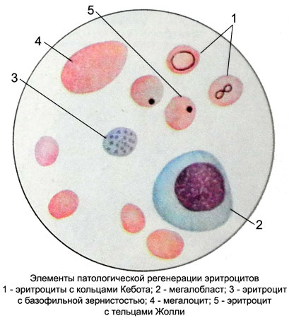
Red blood cells - Elements of pathological regeneration
The elements are pathological regeneration megaloblasts, megalocitы, Jolly corpuscles, Ring Kebota, basophilic stippling of red blood cells.
Taurus Jolly
Taurus Jolly - Small round violet-red the inclusion of 1-2 microns, occurring two or three in one erythrocyte. They are the remnants of the core megaloblasts, delay in erythrocyte in connection with violation of megaloblasts release from the nucleus in the bone marrow. Under normal conditions, these corpuscles can only be detected in the embryo and in the blood of newborns. In the later period, the calf Jolly appear in the peripheral blood in some types of anemia, poisoning hemolytic poisons, and after splenectomy.
Rings Kebota
Rings Kebota represent, apparently, the remains of the nuclear envelope. They look ringlet, Eight or treble clef, turn red. There are only in pathology, mainly at B12– and folic acid deficiency anemia.
Basophilic stippling of red blood cells
Basophilic stippling of red blood cells It observed in severe forms of anemia and toxic states (eg, lead poisoning). When stained with Romanovsky basophilic stippling of red blood cells in blue. It indicates toxic damage of bone marrow and therefore has an adverse prognostic value. Basophil granules, apparently, are the remnants of basophilic substance, similar D- tnkulotsytam.
Other inclusions in erythrocytes
Of the other impurities in the erythrocytes should be pointed graininess, contains non-hemoglobin, readily removable iron (hemosiderin, ferritin). Red blood cells with such inclusions named siderocitami and recognized as precursors of mature erythrocytes. They include ferritin in the form of fine granules (0,5-1.5 M).
Their number in peripheral blood of healthy humans is 0.5-0.8 %. Occurrence siderotsitov in more indicates violation of the synthesis of hemoglobin, that there is at poisoning by lead salts, in some hemolytic anemia and hypochromic sideroblasticheskoy, when less pernicious anemia and thalassemia. Sometimes the increased amount siderotsitov appears in multiple blood transfusions or intravenous administration of iron preparations.
If peripheral blood siderotsity found mainly in pathological conditions, in the bone marrow and are normally sideroblasts - nucleated cells erythrogenesis, in the cytoplasm, which are identified in the special color of iron pellets. Normally, the number of sideroblasts in the bone marrow is 20-40 % bone marrow erythroblasts; iron deficiency anemia when their number is reduced to 2- 5 %. To identify siderophilic granules in normocytes and erythrocytes using special staining techniques, in which the cytoplasm and nucleus of cells painted pink, siderophile and granules - in blue.
Sometimes it may be found in erythrocytes Schuffner's grain or Maurer's clefts (malaria), calf Heinz-Ehrlich (considered the first sign of the coming of hemolysis and toxic damage of blood), nuclear speck Weidenreich (a sign of profound disintegration of the nucleus), However, the origin of these inclusions and their diagnostic value still poorly.

