Red blood cells - red blood cells Degenerative changes
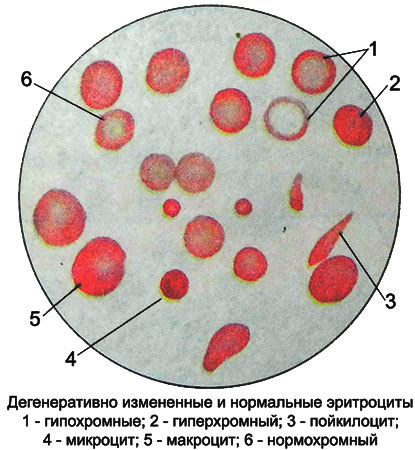
Degenerative changes of red blood cells in pathological conditions may relate to their values, shape and color.
Anisocytosis
Changes in the value of red blood cells - anisocytosis.
Erythrocytes diameter less 6,5 microns are called mikrocitami, and the state, where they predominate - mikrotsitozom (observed in iron deficiency). Erythrocytes diameter greater than 8 um called makrocitami, and the state of their predominance - makrotsitozom. As a physiological phenomenon is observed in newborns and fades to two months of age, as pathological - noted in the disorder of the hematopoietic function of the liver, pernicious anemia, anemia in pregnant women, Cancer, polyps of the stomach, etc..
Erythrocytes diameter greater than 12 microns are called megalocitami. They generally oval, hyperchromatic, They have biconcave (because of the large thickness of no central illumination). Found with a deficiency in the body methylcobalamin - kobamidnogo coenzyme (B12-cofactors) or folic acid. In some cases, (in severe anemia) there are very small fragments the size of red blood cells 2-3 microns - shizocitы.
Anisocytosis is an early sign of anemia. Isolated anisocytosis without other morphological changes in red blood cells observed in mild forms of anemia.
A more accurate picture of the distribution of red blood cells in size can be obtained by measuring its diameter and construction eritrotsitometricheskoy Price-Jones curve. This curve allows you to identify minor quantitative changes in the value of red blood cells and track them at various anemias. The shift of the curve to the left indicates microcytosis, right - of macrocytosis, a broad peak of the curve indicates the presence of anisocytosis.
Poikilocytosis were
Changes in the shape of red blood cells - poikilocytosis were. Characterized by, which in severe forms of anemia red blood cells become elongated, pear-shaped, sharp-edged. Poikilocytosis were - A key feature of the degenerative changes of erythrocytes. Unlike anisocytosis, it develops with severe anemia and is a poor prognostic sign.
When hypochromic anemia due to the decrease in hemoglobin in the red blood cells is reduced and their thickness - formed planotsity (platicity, leptocitы).
Hemolytic anemia increases red blood cell thickness, but they remain biconcave - appear spherocytes. Being exposed to damage in the sinuses of the spleen, they decrease and turn into microspherocytes.
Mikrosferotsitoz
Microspherocytosis - a key feature of hematology hemolytic anemia microspherocytic (disease Minkowski-Chauffard).
Some types of hemolytic anemia red blood cells appear oval - ovalocytes, when sickle cell anemia - drepanotsity, or sickle-shaped red blood cells.
Mishenevidnye cells
Mishenevidnye (kokardnye, Targeted or) cell They represent erythrocytes, in which the hemoglobin is not only along the periphery, but in the middle and; found in thalassemia and other types of anemia (iron), as well as certain liver diseases and in healthy individuals.
If hemolytic conditions can be observed bitargetnye cells, who between central and peripheral portions of hemoglobin are colorless ring. Red blood cells with the unpainted portion in the center (shaped like a mouth) are called stomatocytes.

