Epithelial tissue
Pokrovnıy epithelium
Odnosloynıy epithelium
In the description of a single-layer single-row epithelium, the term "single-row" often falls. Depending on the shape of the cells (эpiteliotsitov) distinguish:
- Flat odnosloynıy epithelium;
- Kwbïçeskïy odnosloynıy epithelium;
- Cylindrical, or odnosloynıy prismatic epithelium.
Odnosloynıy ploskïy epithelium, or mesothelium, lines the pleura, peritoneum and pericardium, It prevents the formation of adhesions between the abdominal and thoracic cavities. When viewed from above the mesothelium cells have a polygonal shape and jagged edges, transverse sections are flat. The number of cores in them varies from one to three.
Dual-cells are formed as a result of incomplete mitosis and amitosis. Electron microscopy can detect the presence of cells on top of microvilli, which significantly increases the surface mesothelium. When the pathological process, such as pleurisy, pericarditis, mesothelia can occur through the intensive release of fluid in the body cavity. With the defeat of the serous membrane of the cells of the mesothelium are reduced, moving away from each other, rounded and are easily separated from the basement membrane.
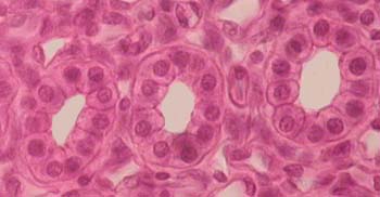
Odnosloynıy kwbïçeskïy epithelium lining tubules in the nephrons of the kidneys, small branching excretory ducts of many glands (liver, pancreatic cancer and so on.). Height and width of a cubic epithelium cells often are about the same. At the center of the cell nucleus is round.

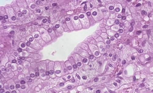
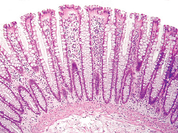
Odnosloynıy cïlïndrïçeskïy epithelium lines the cavity of the stomach, small and large intestines, gallbladder, excretory ducts of the liver and pancreas, and certain forms the walls of tubules of nephrons, etc.. It is a layer of cells of cylindrical shape, disposed on a basal membrane in a single layer. The height of epithelial cells more than their width, and they all have the same shape, therefore their core lie on one level, par.

The organs, where it is constantly and intensely committed the processes of absorption (gut, gallbladder), epithelial cells have a suction rim, which consists of a large number of well-developed microvilli. These cells are called limbic. The edges also contains enzymes, break down complex substances to simpler compounds, can penetrate tsitolemmy (a skin cell).

A feature of a single layer of columnar epithelium, stomach lining,is the ability of cells to secrete mucus. This is called the epithelium of the mucous. Slime, produced by the epithelium, It protects the gastric mucosa from the mechanical, chemical and thermal damage.
Odnosloynıy Multi mercatelnıy cïlïndrïçeskïy epithelium xarakterïzwetsya nalïçïem mercatelnıx cilia, lines the nasal cavity, trachea, ʙronxi, the fallopian tubes. The movement of the cilia along with other factors contribute to the movement of eggs in the fallopian tubes, bronchial - dust particles from the exhaled air in the nasal cavity.
Goblet cells. The single layer of cylindrical epithelium of the small and large intestines meet cells, shaped glasses and secreting mucus, which protects the epithelium from mechanical and chemical action.
Mnogosloynıy epithelium
Mnogosloynıy epithelium There are three kinds of:
- Stratum;
- Neorogovevayuschy;
- Transition.
The epithelium of the first two types of skin covers, Cornea, lines the mouth, esophagus, vagina and urethra; perexodnıy epithelium - poçeçnıe loxankï, ureters, urinary bladder.
The regeneration of the epithelium
Surface epithelium is constantly exposed to the external environment. Through it carried out an intensive exchange of substances between the organism and the environment. Therefore, epithelial cells die quickly. It is estimated, that only the surface of the oral mucosa of a healthy person every 5 min exfoliated over 5-105 epithelial cells.
Epithelial regeneration occurs by mitosis of epithelial cells. Most of the single-layer epithelial cells able to divide, and in stratified epithelium such ability have only basal cells and some thorny layers.
Reparative regeneration of the epithelium It is going through intensive cell multiplication wound edges, gradually closing in on the location of the defect. In the future as a result of ongoing cell proliferation of epithelial layer thickness in the area of the wound increases, while it occurs maturation and differentiation of cells, purchasing structure, inherent in this type of epithelial cells. Of great importance for the process of regeneration of the epithelium has a status of the underlying connective tissue. Epithelialization of the wound occurs only after filling her young, rich in blood vessels, connective (granulation) cloth.
Glandular epithelium
Glandular epithelium consists of glandular, or secretory, cell - glandulotsitov. These cells synthesize and secrete specific products (Secrets) on the skin surface, the mucous membranes and in the cavity or internal organs into the blood and lymph.
The glands in the human body perform secretory function, It is either independent bodies (pancreas, shtitovidnaya, major salivary glands, etc.) or their elements (gland fundus). Most glands - derived epithelial, and only some of them are of a different origin (eg, medulla of the adrenal gland develops from nerve tissue).
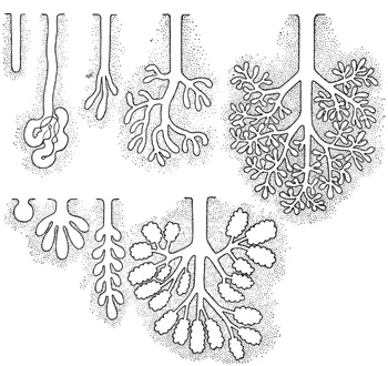
According to the structure distinguish simple (with nevetvâŝimsâ vyvodnym flow) and complex (with razvetvlennym vyvodnym flow) glands and in functions - endocrine gland, or endocrine, and exocrine, or exocrine.

Endocrine glands are gipofiz, pineal body, shtitovidnaya, parashtitovidnaya, thymus, gonads, adrenal glands and pancreatic islets. Exocrine glands produce secret, released into the environment-on skin surface or in a cavity, lined with epithelium (gastric cavity, intestines, etc.. d.). They participate in executing the function body, elements of which are (eg, cancer of the alimentary canal are involved in digestion). Exocrine glands are different from each other Location, structure, type and composition of the secretion secret.
Most of exocrine glands – multicellular Education, except the goblet cells (the only species of unicellular exocrine glands in the human body). Goblet cells located within the epithelial layer, produce and secrete mucus on the surface epithelium, protects it from damage. These cells have extended tip, which stores the secret, and the narrow base with a nucleus and organelles. The rest of the exocrine glands - multicellular ekzoepitelialnye (located outside the epithelial layer) education, which distinguish secretory, or end, Front and excretory duct.
Secretory Department It consists of secretory, or glandular, cell, generating a secret.
In some glands, Derivatives stratified epithelium, besides the secretory epithelial cells occur, retractility. Cutting, they compress the secretory department, and thus facilitate the release of his secret.
Cells secretory departments - glandulotsity - often lie in one layer on a basal membrane, but may be located in several layers, for example in the sebaceous glands. Their form varies depending on the phase of secretion. Cores are usually large, irregular shape, with large nucleoli.
The cells, generating the secret nature of the protein (eg, digestive enzymes), particularly well developed granular endoplasmic reticulum, and cells, producing lipids and steroids, better expressed nezernistaya endoplasmic reticulum. Well developed plate complex, directly related to the processes of secretion.
Numerous mitochondria are concentrated in areas most active cells, t. it is. there, which accumulates secret. In the cytoplasm of glandular cells are found various kinds of inclusions: protein grains, drops of fat and glycogen clumps. Their number depends on the phase of secretion. Often between the side surfaces of cells are intracellular secretory capillaries. Citolemma, limiting their clearance, forms numerous microvilli.
In many glands clearly visible polar differentiation of cells, due to the orientation of the secretory process,- Synthesis secret, its accumulation and secretion in the lumen of the end section in the flow direction from the base to the apex. In this connection, the cells are located in the grounds of the kernel and ergastoplasm, and the tops of the mesh device is intracellular.
In education, there are several secret successive phases:
- Uptake for the synthesis of secretion;
- The synthesis and accumulation of secretions;
- Secretions and restore the structure of glandular cells.
The release of secret occurs periodically, in connection with which there are regular changes of glandular cells.
Depending on the method of allocation of a secret distinguish merokrinovy, apocrine secretion and holocrine types.
When merokrinovom type secretion (the most widespread in the body) glandulotsity fully retain their structure, secret exits the cell into the cavity through the hole in the gland tsitolemmy either by diffusion through tsitolemmy without compromising its integrity.
When apocrine type of secretion grandulotsity partially destroyed, and at the tip of the secret cells separated. This type is characteristic of the secretion of milk and some sweat glands.
Holocrine type of secretion It leads to the complete destruction of glandulotsitov, which are part of the secret with them synthesized substances. The man on the type holocrine only secrete sebaceous glands of the skin. With this type of secretion of glandular cells restoration of the structure is due to the intensive propagation and differentiation of specific cell undifferentiated.
The secret of exocrine glands can be a protein, mucous, protein and mucous, greasy, also referred to as the corresponding cancer. In mixed cell glands occur two kinds: Some produce protein, others - mucous secretion.
Excretory ducts of exocrine glands consist of cells, non-secretory capacity. In some glands (Salivary, sweat) excretory ducts cells may participate in the process of secretion. The glands, developed from stratified epithelium, wall outlet ducts are lined with stratified epithelium, and glands, derivatives are single-layer epithelium - single layer.
