Elements of bone and cartilage tissue
Bone
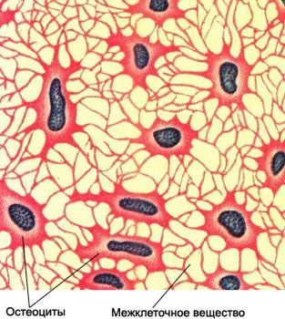
To the cells are bone osteoblasts and osteoclasts. Osteoblasts form bone, consisting of mature cells - osteocytes and mucoid substance - osseomukoida, to postpone salts of calcium therein, tight-knit cells. Osteocytes in cytological preparations missing.
Punctate, derived from the hearth, or destruction of the tumor, subjected to macroscopic study and description, then, preparations for microscopic examination - first native (unpainted), and then stained. For proper evaluation of cytological data necessary to differentiate osteoblasts and osteoclasts.
Osteoblasts - Small and medium-sized intensely colored cells, substantially slightly elongated with one or two short processes.

Individual cells dramatically elongated and resemble fibroblasts. Osteoblasts can be oval, and irregularly circular shape. Cores of hyperchromic, Small, the same magnitude, arranged eccentrically. Chromatin structure nechetkaya, isolated nuclei contain nucleoli. Basophilic cytoplasm, relatively wide (primarily on one side of the nucleus), often fibrous, but it can be grainy or soft foam. The boundaries are clearly delineated cells.
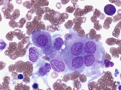
Osteoclasts - Giant multi Process cell, the length and shape of the appendages may vary considerably.

Some have short osteoclasts, like curved spines, others - and even longer fissile. Kernels light, equal size, mainly oval, which are located randomly, most Central. The number of cells varies from district 3 to 20 and more. Xromatin melkozernistыj, compact. The nuclei are isolated nucleoli. Abundant cytoplasm of osteoclasts, basophilic, often with a pinkish tinge. Characteristic for this type of cell is the presence in the cytoplasm of small grain oksifilioy, which can raise the whole of densely cytoplasm, causing her reddish-pink color. In a number of cells and little graininess or it is distributed over the entire area of the cell, or is it just sprouts.
Osteoclasts must be differentiated with giant cells Pirogov-Langhans giant cells and foreign bodies. The pathognomonic for tuberculosis giant multinucleated cells Pirogov-Langhans core characteristically located on the periphery of the cytoplasm, and in the center of their cells no. The shape of the nuclei oval or oblong-bean. The giant cells of foreign bodies contains various amounts rounded nuclei, located in the center of the cells. Their cytoplasm sharply basophilic. The cells of foreign bodies found in punctate inflammatory processes.
Cartilage tissue
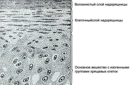
Cartilage tissue consists of cartilage cells and a large amount of intercellular substance. Punctate cartilage has a semi-transparent view of the cylinder. In preparations, made with punctate, It found a small amount of cartilage cells and intercellular substance.

Cartilage cells. The native specimens seen more clearly, since poorly perceive color. Their shape is round, oval or slightly elongated. Tsitolemmy clearly visible. Nuclei are round or oval with gently-point chromatin, occupy a central position. Occasionally there are dual cell. The cytoplasm contains a lot of liquid, so badly stained. Cartilage cells located in the intercellular substance.
Intercellular substance in cytologic specimen looks structureless mass, painted in pink or bluish color. It comprising hondrinovyh (collagen) fibers and ground substance.
Cartilage basic substance - Jelly mass, filling the space between cells and collagenous fibers. It participates in the exchange between the blood cells and. The structure of the basic substance include macromolecular glycosaminoglycans (hyaluronic and chondroitin acid, Heparin, which are firmly bound to proteins). Glycosaminoglycans are dependent on the number of texture, morphological properties and functional properties of the base material. Painted basic substance alkaline dyes in blue tone, and collagen fibers - acidic dyes in pink. In cytological preparations predominant color of the component, which is more than in the intercellular substance.
Myxomatous cells in unstained cytologic preparations can be easily mistaken for artifacts, since nucleus therein are determined with difficulty and not all cells. Sometimes, instead of the nuclei detected small clusters of shiny grains. The small cells stained preparations, different shapes, poorly perceived dye. The nuclei of many cells do not stain, in such cases, the cells look like cytoplasmic structures of various shapes. Stained nuclei small, round or oval, with fuzzy edges.
Myxomatous cells are arranged in isolation or in small clusters, are interconnected processes. These cells are characteristic of the myxoma, and mucilaginized chondroma, fibroma, chondrosarcomas, etc.. Especially a lot of myxomatous cells in cytological preparations at miksosarkomah.

