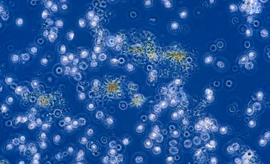
Elements of sludge, found in pathological urine – Microscopic examination of urine
Leucine and tyrosine crystals in the urine sediment usually observed at the same time.
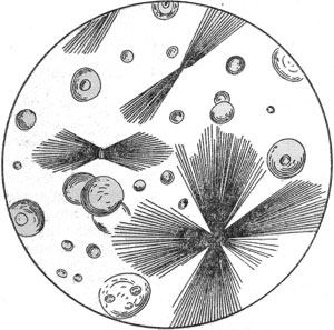
Unlike other crystals, they do not occur in normal urine. They can be found in acute yellow atrophy of the liver, phosphorus poisoning, leukemia and other pathological conditions.
The crystals of leucine form a yellowish-brown or greenish-yellow balls of various sizes with a radiant and concentric striation, reminiscent of a cross-section of a tree. Often on the balls of larger diameter bugroobrazno superimposed smaller.
Crystals tyrosine made up of the finest, brilliant needles, forming a gentle yellowish beams and stars. Leucine is not dissolved in acetic acid, ether, acetone and alcohol. Sometimes it can be mistaken for a drop of fat. It should be borne, that under the influence of ether fat soluble, and leucine remains unchanged. From ammonium urate leucine is characterized by a circular and radiant striation, and the absence of spines on the periphery. Tyrosine crystals can sometimes be confused with neutral phosphate crystals. Latest, Unlike tyrosine, easily dissolved in acetic acid. Tyrosine is soluble in mineral acids and alkalis.
Cystine macroscopically looks greyish-white mass, which under the microscope are correct colorless transparent hexagonal plates, lying next to one another or.
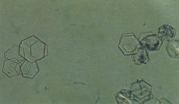
Cystine crystals are insoluble in water, alcohol, ether, acetone, acetic acid, but soluble in mineral acids, ammonia and alkali, and different than some similar forms of uric acid crystals, which are soluble in alkalis, but not dissolve in ammonia.
Cystine appears when hereditary cystinuria. This usually highlights cloudy greenish urine- yellow color, generally alkaline or very slightly acidic. Abundant grayish-white precipitate is composed of crystals and often as many as cystine aggregates of various sizes. As a result of trauma during the passage of salts in the urine can be detected and the admixture of blood. Because of the rarity of the disease and diagnosis of responsibility for carrying out chemical reactions it is mandatory.
Ksantin in the urine sediment is rare. It has the form of colorless rhombs, more reminiscent of uric acid crystals, but, in contrast to the latter, It gives a negative response and mureksidnuyu equally well soluble in alkalis, ammonia and hydrochloric acid, whereas crystals of uric acid or in ammonia, or insoluble in acids.
Cholesterol crystals sometimes found in the urine in the amyloid degeneration of the kidneys and lipoid, echinococcosis urinary tract neoplasms of the urinary and genital organs, mainly cancer and renal abscess. The crystals have the form of colorless small and large plates with cut corners and a step-like ledges. They are located either alone or superimposed on each other. The alkali and acid insoluble crystals, but readily soluble in chloroform, ether and hot alcohol.
Fat and lipids in urine. Fat has the form of highly refractive light droplets and grains with sharply defined dark edges. They may be in the free liquid or layered on shaped elements. Drops of fat cells often filled with so, that they become invisible core, and the cells themselves take the form of rounded formations, entirely composed of fat droplets. Drops of fat also found in clusters in the fatty degeneration and decay of tissue. Fat can occur in the form of incidental impurities from the outside. Neutral oil is dissolved in chloroform, air and stained with Sudan III in the orange-red color, and 1 % osmic acid solution - black.
Morphologically identical fat droplets in the urine in some cases may be a neutral fat, Other - Lipids.
Lipids possess dual refrangibility, which can be detected by polarized light, using the two Nicols. One prism (polarizer) inserted into the aperture of the microscope, second (analyzer) nasazhivayut of eyepiece. If prisms are arranged parallel to each other, Field of view is the usual form; if the analyzer is rotated by 90 °, the field of view becomes dark. It can be seen only in lipids as bright bodies. Lipid droplets look like black cross with four glowing segments, neutral fat droplets thus invisible. Twofold refrangibility also have crystals of fatty acids, but morphologically they are easily distinguishable from lipids. Crystals fatty acids have the form of slightly curved needles, collected into bundles. They are readily soluble in ether and chloroform.
Fat in the urine appears in nephrotic syndrome, and other diseases.
Gematoidin - Crystalline pigment (derivative hemosiderin), It occurs in the form of orthorhombic needles or plates, collected into bundles and sprocket, or in the form of piles and clumps, consisting of small grains. Depending on the size of the crystals and their thickness hematoidin colored in varying shades from golden yellow to brownish-orange.
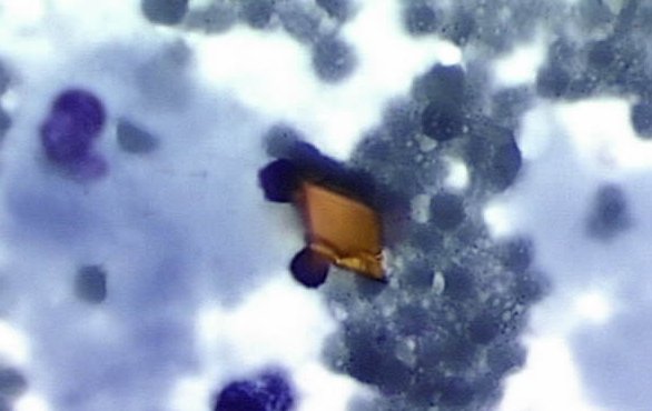
Hematoidin does not contain iron and is formed in the foci of necrosis without oxygen. Inside the cells are not found crystals gematoidina. Have urine gematoidina observed in calculous pyelitis, renal abscess, prostate. Hematoidin can also be seen in the small pieces of necrotic tumors of the cervix, Bladder and kidney. With nitric acid gives fast disappearing blue color.
Hemosiderin found in urine sediment as amorphous masses, which are deposited on all elements of the urinary sediment, giving them a brownish tint.
Bilirubin It appears in the urine as icteric needles tan, either alone or part lying crosswise one above the other, part of the beam; some of them are slightly bent. Bilirubin crystals are deposited on the surface of white blood cells, epithelial cells. Besides, bilirubin occurs as amorphous pigment grains. It gives nitric acid green color, soluble in alkalies and chloroform.
