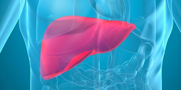
Hydatid disease of liver – treatment of disease. Symptoms and prevention of liver hydatid cyst diseases

Hydatid disease of liver – What is this disease? Hydatid disease of liver (LAT. echinococcosis of the liver)- human helminth infections, characterized by the formation in the liver, parasitic tumor formations-cysts.
Hydatid disease of liver – The cause of the
Ways of infection:
- – eating unwashed vegetables, fruit, berries;
- – raw water from natural reservoirs;
- – by contact with contaminated soil;
- – from infected wild or domestic animals.
Symptoms of liver hydatidosis
For cystic hydatidosis is characterized by the formation of one or more cysts, often, they are localized in the right lobe of the liver, and that is the, later, cause of pain in the liver and other internal organs of neighbouring. Pain sensations arise from mechanical impact cysts on data field. The parasite also has toxic effect on the entire organism, allocating in blood products own vital functions. Jehinokokkovaja cyst has a dense layered shell thickness to 5 mm, on the form, the cyst is reminiscent of bubble.
Most often, patients in the early stages of hydatidosis is missing, any, clinical manifestations, only with an increase in the size of the cyst in a patient appear dull persistent pain in the right hypochondrium and epigastrium, as well as in the lower divisions of thorax. By palpation of the liver detected tumorous formation.
Depending on the location of the cyst, There are three types of hydatidosis: front, descending, ascending. With front-type front liver region significantly increased, when descending tumorous type education has closer to abdominal cavity, When ascending the type of cyst has a domed shape.
Hydatid disease of liver – Diagnostics
In order to diagnose liver hydatidosis using x-rays. Using x-rays can detect the presence of cysts in the liver, determine the shape of a wall, as well as find indirect signs of cysts, such as: high location or deformation of the dome of the diaphragm, the offset arrangement of loops of small intestine, stomach displacement.
Method of selective angiography, perhaps, determine the degree of contrast material accumulation between the cyst and the shell of fibrous capsule .
Using the radioisotope scanning, You can define a rounded defect drug accumulation in the liver.
When tubal interruption using laparoscopy method, defines the location of the parasitic cysts in the liver.
An important method of diagnosis, are biological tests: intraskin tryout Casoni, reaction of agglutination with LaTeX, reaction of the indirect gemaggljutnnaiii.
More important tryout Anfilogov, that shows an increase in the number of eosinophils after palpation for cysts, It is valid only in case of, If the parasite is alive, After the death of the parasite this symptom is not present.
Hydatid disease of liver – Types of disease
On the structure of the entities, There are two types of hydatidosis:
- – alveolar or multilocular echinococcosis , is characterized by an extensive degree of liver;
- – Cystic or multi-Chamber Hydatid disease, resembles a bubble, around which formed a complex layered shell, that hosts the derived capsules.
On location, There are three types of hydatidosis:
- – front, with the progression of this type hydatidosis, dimensions of the liver significantly increases, it becomes available by palpation;
- – top-down type of hydatidosis, lodging the report closer to the abdomen, his consistency is elastic;
- – bottom-up type, on semiology recalls ekssoudativei Pleurisy, on radiographs, has the appearance of a high stress situation of the dome of the diaphragm.
Hydatid disease of liver – Actions of the patient
Patients should urgently appeal to the doctor for diagnosis and treatment.
Treatment of liver hydatidosis
Treatment of cystic, and alveolar hydatidosis, often quite expensive and prolonged.
There are four main options for treating cystic hydatidosis type:
1. Utilising PAIR (needling, Aspiration, injection, reaspiracija).
2. Surgical treatment method.
3. Application of anti-infective drugs.
4. Monitoring method.
Tactics of treatment and therapy should be based on the results of diagnostic studies (kistyt ultrasound, etc.). You must also take into account the stage of disease.
When alveolar jehinokokkoze, important is early diagnosis and radical surgery with holding, subsequent, anti-infectious therapy. If you defeat the limited nature, the surgical treatment leads to recovery of the patient.
In the case of a running stage disease palliative surgery, held together with incomplete anti-infectious therapy or without it, often leads to recurrent disease.
Hydatid disease of liver – Complications
With the development of complications of hydatidosis-inflammatory conditions and, later, rupture cyst, the clinical picture can be complex. Characteristic symptoms in nagnoivshijsja jehinokokke, It is: fever, pain in the liver, neutrophilic leukocytosis, ESR acceleration, anjeozinofilija, toxic neutrophils ' granularity.
A breakthrough in the case of cysts in the bile path and/or blood vessels, patient's itch, hives on skin layers, sometimes anaphylactic shock. May develop peritonitis, at the opening of nagnoivshegosja cyst in the cavity the peritoneum. Gap jehinokokkovoj cysts in the Airways can cause secondary pulmonary hydatidosis.
Prevention of hepatic hydatidosis
For personal prevention liver hydatidosis, You should limit contact with dogs, Games , wash hands thoroughly after each contact with animals. Be sure to wash your hands before you eat after working in the garden or on a kitchen garden, After collecting mushrooms in the forest. Do not take in food not washed wild fruits, don't drink don't drink water from natural sources.
Preventive measures also include veterinary-medical activities in detecting hydatidosis, They mainly aimed at identifying and eliminating the source of the infestation.
As an additional measure of prevention addresses the vaccination of sheep against hydatidosis.
