Benign breast tumors
Papilloma ducts
Papilloma of the milk ducts is usually localized in the large milk ducts of the breast around the nipple; fine, vnutriprogokovye papillomatoznye sprawl, characteristic mastitis, a duct papilloma do not apply.
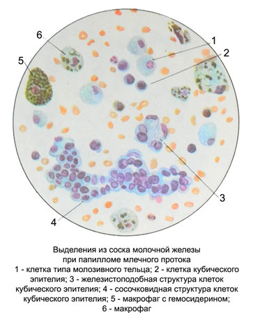
For papilloma ducts characterized bloody discharge from the nipple character. In the preparations of selections on the background of erythrocytes modified and unmodified, macrophages with hemosiderin, single flattened cells and histiocytes found epithelium ducts are able to moderate proliferation of cell complexes characteristic elongated shape, similar to microscopic papillomas.

Papillomavirus can have multiple branching papillary form, which makes the entire structure intricate contours. Usually, papilloma composed entirely of epithelial cells, Only occasionally complexes like with axial filament, formed connective elements.
Mleçnıx duct epithelium can also be placed separately and in the form of small round or irregularly shaped structures zhelezistopodobnyh, consisting of a small, intensely stained cells with the same enlarged hyperchromatic, having smooth contours of the nuclei. The cytoplasm of many cells vacuolated. Cages, lying separately on the edges complexes, Larger, bright, as if swollen. The outer layer of cytoplasm have sealed. Complexes of closely contiguous cells stained intensely, a small increase dramatically stand out against the drug, and under a lot of ill-viewed. On the periphery of such complexes cells often arranged in rows. The boundaries between the cells often indistinguishable. Complexes of the larger cells by a pronounced proliferation stained less intensively. The preparation of the complexes may be cell-type cells colostric, which differ in larger size, lack of regular rows, disorderly dispersion of cores. Besides, complexes from cells colostric type cells are present only in those preparations, in which a lot of disparate, clear, There is no doubt the cells of the same type.
When papilloma ducts may occur premalignant proliferation, which is characterized by the appearance of signs of atypia in the epithelial cells and the presence of secretions from the nipple of the breast malignant cells. In violation of the outflow of such cells can not enter the discharge from the nipple. A sign of malignancy is the presence of a massive circular complexes or cell papillary structures with random arrangement of epithelial cells.
Atypia cellular elements It may manifest as a change in the size and shape of the cells, and in violation of the size and shape of the nuclei, Change cores- but-cytoplasmic ratio.
Leaf-breast tumor – Intraductal fibroadenoma
Leaf-breast tumor (intraductal fibroadenoma) - A special kind of fibroadenomas, which is characterized by rapid growth and can reach a diameter 20 cm. There are three types of leaf-tumor:
- classical stromal cells without atypia;
- predsarkomatoznuyu, predsarkomatoznymi characterized by changes of the stroma, proliferation of fibroblasts, fibrotsitov, the presence of cartilage, bone, adipose tissue and vascular atypia individual cells;
- sarcomatous, whose stroma different from stromal sarcoma. Epithelial tumors these elements are in a state of moderate proliferation and rarely in cancer metaplaziruyut (carcinosarcomas).
Cytological studies often exposed scrapings and punctate; discharge from the nipple of the breast are rare.
Scrapings from the cut of the tumor moderate, seromucous nature, gray, sometimes with a yellowish or reddish tinge, with small scraps of fabric.
Punctate moderate, sanioserous nature, with small scraps of fabric. With rapid growth of the tumor, accompanied by skin ulceration, discharge from the nipple appear sanioserous nature.
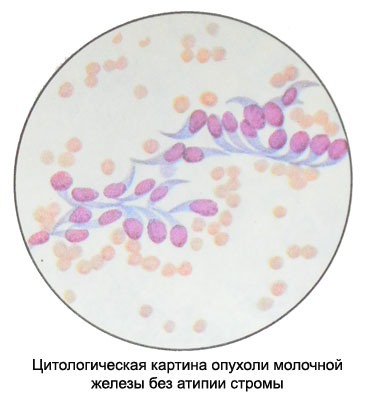
Leaf-tumor stromal cells without atypia
Background preparation is represented by fine-grained slizevidnymi oxyphilic tyazhistymi or structureless masses. A large number of fibroblasts, fibroblasts and epithelial cells. Epithelial cells of small, intensely colored, cubic or prismatic shape with identical round nuclei, nucleoli are not visible. Epithelial cells are arranged separately and in groups, They are often closely linked with fibroblasts and fibrocytes, beam forming and star clusters.
Fibroblasts and fibrocytes are placed in isolation, groups or strands.

The proportion of epithelial elements, fibroblasts and fibrocytes may be different. When a small amount of the preparation of fibroblasts differentiate tumor from leaf-nelistovidnoy fibroadenoma and mastitis glandular fails.
A feature of leaf-tumor This type is the presence of foam cell type lipofagov, gistiocitov, myxomatous myoepitheliocytes, which is not characteristic of other fibroadenomas.
When predsarkomatoznoy leaf-tumor You can detect fat, myxomatous, cartilage cells, osteoblasts and osteoclasts, and giant cells with scalloped edges, bizarre, with signs mucilaginized. Occasionally there are atypical cells with large hyperchromatic nuclei. There mitotic figures.
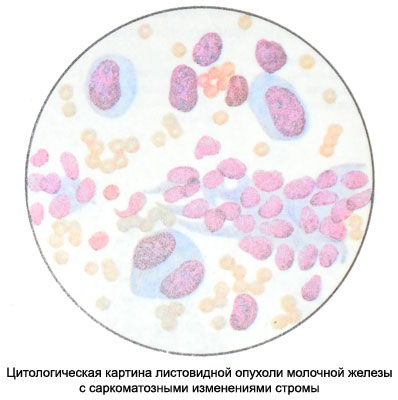
Sarcomatous tumor of the leaf – Lystovydnaya tsystosarkoma
Against the backdrop of blood and necrotic masses (often mucilaginized) found polymorphic atypical cells of connective stroma, while dominated by bizarre fibroblast-like cells with a sharp increase in the nuclei and nucleoli hypertrophied, settling loosely. Many cells in the pathological state of mitosis. When malignancy cartilage and bone components of the tumor detected atypical osteoblasts, osteoclasts and cartilage cells. Epithelial cells are typically in small numbers, with degenerative changes and sometimes with signs of malignancy.

Against the background of leaf-tumor cancer or mixed tumor (carcinosarcomas) It is developing very rarely. Grandular proliferous cyst is different from other types of sarcoma presence of epithelial component.
