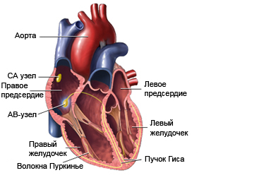
Arrhythmia
Description of the arrhythmia
Arrhythmia – abnormal heartbeat. Types of arrhythmias include:
- Too slow heartbeat (bradycardia);
- Too rapid heartbeat (tachycardia);
- Additional (unnecessary) heart beats;
- Skipped heart beats;
- Strikes in abnormal areas of the heart.

Causes of arrhythmia
Arrhythmia can be caused by:
- Natural stimulant heartbeat (atrionector) sets the wrong speed or rhythm;
- Normal heartbeat is interrupted for no apparent reason;
- Another part of the heart begins to perform the functions of sinoatrial node.
Risk factors
Factors, that increase the likelihood of arrhythmias:
- Excess caffeine;
- Stress;
- Tobacco;
- Alcohol consumption;
- Taking some diet pills or protivootechnyh funds;
- Cocaine use;
- Some antidepressants;
- An overdose of Digitalis;
- Endocrine disorders, such as the thyroid and adrenal problems;
- Anemia;
- High blood pressure;
- Coronary artery disease;
- Problems of the heart valves;
- Damage to the heart muscle after a heart attack;
- Revmokardit;
- Cardiomyopathy;
- Diabetes;
- Liver disease;
- Typhoid fever;
- Gipotermiя;
- Electric shock or lightning;
- Complications after partial drowning.
Arrhythmia symptoms
Sometimes the arrhythmia can occur without any symptoms. But some species may cause noticeable symptoms, such as:
- Fainting;
- Dizziness;
- Palpitations;
- The feeling of missed or extra heart beats;
- Weakness;
- Fatigue;
- Breathlessness;
- Chest pain.
Diagnosis of arrhythmia
The doctor asks about the symptoms and medical history, performs a physical exam. Usually the doctor listens to the heart of the stethoscope and may appoint the following surveys:
- Blood tests – to look for certain markers in the blood, that help the doctor determine, What happens to the heart;
- Urine – to look for certain markers in the urine, that help the doctor determine, What happens to the heart;
- Electrocardiogram (ECG) – record heart activity by measuring electrical current, passing through the heart muscle;
- Echocardiogram – uses high-frequency sound waves (ultrasound), to examine the size, shape and motion of the heart;
- 24-hour Holter monitoring – Portable ECG recorder attached to the patient, to record heart activity during normal daily activities;
- Stress test – record electrical activity of the heart during increased physical activity. Patient, who cannot exercise, can be entered by intravenous means, to simulate the effect of physical exercise on the heart;
- Nuclear scanning – radioactive material is injected into the vein, and note his absorption of cardiac myshcoj. Areas with reduced blood flow, and, Consequently, lower absorption of radioactive material, visible as dark spots in the picture;
- Coronary angiography – after administration of the radiopaque dye into the coronary artery is performed radiograph, that allows the doctor to look for anomalies (narrowing, blood clots) in the arteries and to evaluate heart function;
- Electrophysiological study – in this study, current is passed through the blood vessels by using electrode catheters, attached to different places on the heart. This allows doctors to see, as electrical impulses pass through the heart, and helps diagnose problems conduction and/or tissue identification, that can cause arrhythmia;
- Testing using ortostaticski backgrounds – This test is usually recommended for patients with fainting episodes. The patient lies flat on the table, that swivels to vertical position. During the inclination measured heart rate and blood pressure, as well as other parameters of cardiac activity.
Treatment of arrhythmia
Treatment may include:
Admission medication complications
Medications can help slow down or speed up the heart rate, as well as return the heart to its normal rhythm (cardioversion), Depending on the patient's needs.
Electric or cardioverter defibrillator
These treatments include placing special device on his chest or back. An electric current passes through the chest wall to the heart, and gets the heart rhythm back to normal.
Automatic implantable defibrillator
A miniature defibrillator can be surgically embedded in his chest, to control heart rhythm. If there is already, device automatically tries to restore heart rhythm back to normal.
Artificial pacemaker
The Stimulator is implanted into the chest. It produces electrical impulses, necessary for normal heart rhythm.
Ablative therapy
The area of the heart, who is responsible for the abnormal rhythm can be surgically removed or going through a variety of methods (eg, using krioabljacii), to prevent arrhythmia.
Arrhythmia prevention
To prevent arrhythmia must be:
- Cure disease, that can lead to arrhythmia;
- Avoid substances, that can cause or aggravate arrhythmia, which include:
- Caffeinated drinks;
- Alcohol;
- Tobacco;
- Some medications;
- You should follow the General rekomendacjam for the prevention of cardiovascular disease:
- Support a healthy weight;
- Engage in sports and physical education;
- You must quit smoking;
- You must eat healthy food, which contains little saturated fat and rich in whole grains, fruit, and vegetables;
- Timely treat high blood pressure and/or diabetes.
